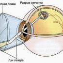
Retinal detachment - a disease requiring special attention. The success of treatment depends primarily on the early diagnosis of the disease. In our article, we consider the methods of diagnosing retinal detail
Content
Diagnosis of retinal detachment
 The early diagnosis of retinal detachment is of paramount importance, since it allows you to start the treatment timely and prevent the inevitable loss of vision.
In the process of man survey with retinal detachment, the following three groups of research methods can be distinguished:
- Standard (routine) methods of ophthalmological examination
- Determination of visual acuity (visometry)
- Study of fields of view (perimetry)
- Measurement of intraocular pressure (tonometry)
- Study of the front segment of the eye (biomicroscopy)
- Inspection of the eye dna with a wide pupil (ophthalmoscopy)
- Study of entopic phenomenov
- Additional (special) research methods appointed if necessary
- Ultrasound in mode
- Electrophysiological research methods (electrical sensitivity threshold, optic nerve lability, critical fission frequency flashes)
- Laboratory research methods (when preparing for hospitalization)
The first group includes the methods of standard ophthalmic surveys. A special role among them belongs to inspection of the eye dove – Ophthalmoscopy. Inspection of the Eye DNA allows you to determine the degree of length of the retinal detachment, detect the retinal breaks, estimate the threat of a threat of a macular area, to determine the probable place of the primary break by the configuration of the detachment and, most importantly, allows you to determine the tactics of surgical treatment.
Ophthalmoscopy – key method when examining patients with retinal detachment.
In the arsenal of the ophthalmologist, there are several techniques for inspection of the ocular bottom (using a contactless lens, using a three-member goldman lens, with the help of a naked ophthalmoscope and others.). Combination of several techniques allows you to get the most complete information about retinal detachment.
 Inspection of the eye dna It is carried out repeatedly, both in the sitting position and in the position lying. This allows the doctor to carefully examine the extreme periphery of the eye bottom, to detect all the gaps and zones of the retinal degenerations, the closure of which is the basis of therapeutic measures during the retinal detachment surgery.
In the group of standard methods, the study of entopic phenomena (mechanophosphen, autoophthalmoscopy phenomenon, etc.).
The study of the mechanophosphate is carried out by pressing the glass stick on the scler in the retinal projection site. At the same time on the side opposite to the pressing, a dark or light spot is felt.
The autoophthatmoscopy phenomenon (T.E. Observation by the patient's own retinal vessels) is carried out by lighting a sclera of the examined eye with a bright point light source. In this case, the patient sees the picture of his own retinal vessels, which are represented in the form of a branch of a tree, «cracks» and «Crossow».
These simple methods allow you to presumably judge the presence or absence of retinal detachment with severe clouds in the lens, hemorrhage into the glades cavity, eliminating the possibility of a detailed examination of the eye dna.
The second group includes special research methods. Thus, an ultrasound study in in-mode is used mainly with severe turbines in a lens and a vitreous body, when ophthalmoscopy capabilities are sharply reduced or its implementation is impossible.
The role of electrophysiological studies is secondary, however, they can be useful in assessing the viability of the retina in the event of an old detachment.
Laboratory tests and a number of other research methods are appointed to the patient hospitalization. These include a general blood and urine test, biochemical blood test, blood test for HIV, syphilis, hepatitis B and C, radiograph of the chest organs and the apparent sinuses of the nose. In addition, the conclusion of the therapist, dentist, LOR doctor, according to indications – other specialists (endocrinologist, nephrologist and others.). All this is done to identify contraindications to surgery (decompensation of common diseases, identification and rehabilitation of chronic infection foci), which may complicate the flow of the postoperative period.
In the case of a fast-moving retinal detachment, when there is a direct threat for a macular region, hospitalization is possible «Cito» (T.E. Essentially, urgently). Then the patient can be hospitalized without the complete above-mentioned set of analyzes and consultations of specialists (only blood tests are necessary). This increases the potential risk of developing complications, but allows you to get the best functional effect due to the timely operation.