Makulyodistrophia - Harbor Dangerous. At the same time, the eyes are not hurt at this, but the consequences of the maculodistrophy can be quite crying. That is why the early diagnosis of this disease is important. Fortunately, it does not represent difficulties. In some cases, you can spend it yourself. Read more about this in this article.
Content
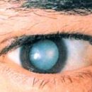
Makula is the central part of the photosensitive layer of the eye - retina. For vision of Makula is very important, at the expense of it we see items that are directly in front of our eyes, t.E. With the help of macula, we read and write. Also Makula gives us the opportunity to distinguish colors.
Makula consists of gentle, photosensitive cells. As a result of some processes, these cells may be damaged, such a condition is called «Makulyodistrophy retina». If the maculodystrophy is developing in old age, it is called «Shenyl maculodystrophy of the retina».
Ophthalmologists classify with maculyodistrophy for two types: wet and dry.
Dry maculodystrophy of the retina occurs more. It develops very slowly, causing the loss of the central field of view. Currently, the doctors still do not know how to treat dry maculodystrophi.
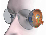
Wet maculodystrophy occurs about 10%. The cause of its development is the formation of new blood vessels behind the retina. New vessels are obtained fragile and permeable for blood, and this leads to the appearance of hemorrhages, the formation of connective tissue and, in the end, to loss of vision. «Wet» Maculyodistrophy develops quickly, in the early stages it can be treated.
Usually maculodystrophy of the retina strikes both eyes, but often one eye begins to lose vision much earlier - in this case, the patient may not notice the beginning of the disease immediately, t.To. One eye compensates for the loss of the second function.
Maculodystrophy does not cause pain and rarely leads to full blindness, t.To. Only the central field of view is affected. Makulyodistrophia is the most frequent reason for the loss of visual acuity in people over 60 years old, as has already been said.
Usually the diagnosis of difficulties does not represent, t.To. Symptoms are characteristic.
Some signs of maculodystrophy can manifest themselves until the moment of direct impairment of view, but only the study of the eye will help to identify them in a timely manner.
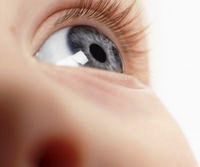
Lounge can be detected with a normal verification procedure when patients burst special drops to expand pupils so that the doctor considers the back wall of the retina. In addition, the doctor will check the ability to see at different distances, with different lighting in the most suitable corrective glasses. In some cases, he can also spend a test on the ability of the eye to distinguish the shades of colors, t. E. Check the status of mesh colums. A complete examination of the eyeball will allow to identify the accompanying diseases, such as cataract and glaucoma.
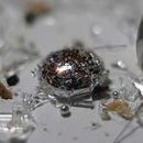
The doctor may suspect the dry shape of the maculodyfia, finding dubs - small multiple whitish-yellowish formations around the central fossa of the yellow spat or excessive pigmentation, which can be considered in an ophthalmoscope. These changes are early symptoms of maculyodistrophia.
Of course, the maculyodistrophia is not completely cured, but it is important to identify maculyodistrophy at an early stage, when it is still possible to prevent the deterioration of the retina state. It is necessary that people aged 40 to 64 passed a survey every 2-4 years, and those who are for 65, -en.
 If you have a dry maculodystrophy, you can control your condition at home, independently determining whether the disease does not go into a wet form. This observation is easy to organize at home using «Amslera grid», which is a sheet of paper in a cell of 10 x 10 cm with a black point in the middle.
Attach this sheet of paper on the refrigerator door or any other place convenient for you. Check each eye very carefully. If the lines near the point seemed to you wavy, it may indicate the development of wet maculodystrophia.
If you have already passed a laser treatment, then regular checks with «Amslera grids» may prevent you about resuming hemorrhages in vessels. Leaf S «Mesh Amselra» Play at a distance of 12-15 inches from eyes with good lighting. Put on reading glasses and close one eye. Look at the point in the center, while pay attention, whether the lines of the lattice are straight or wavy, faded or dark. Repeat the procedure with another eye.
If some areas of the lattice seemed wavy, faded or dark, report this ophthalmologist.
Do not forget to check the second eye!