Every year, more and more people find out that they have melanoma - one of the most dangerous types of cancer. Early diagnosis is crucial to increase chances to defeat the disease. Therefore, we recommend all people with an increased risk of melanoma to check the skin from a specialist.
Content
Examination on melanoma
If any formation on the skin causes suspicion about melanoma or skin cancer, then a special examination is necessary to confirm or exclude the diagnosis.
At the doctor's
 The doctor will ask you about the symptoms of the disease and risk factors, when for the first time appeared on the skin and was it changed in size and appearance. You can ask you for contacts with known substances causing skin cancer, as well as cases of skin cancer in your family.
During the examination, the doctor will determine the size, shape, color and condition of tissues around education on the skin. In addition, he will find out whether there is no moisture or bleeding from suspicious education.
With the inspection of the whole body, all stains and moles will be studied, which may be related to skin cancer. Limph nodes will also be examined in inguinal and axillary areas, on the neck and especially close to a suspicious focus. An increase in lymph nodes can speak in favor of their defeat of a malignant tumor.
Skin biopsy or take a small section
Biopsy is the take of a small area of any fabric for a subsequent detailed study under the microscope. The procedure is performed under local anesthesia in outpatient conditions. Skin biopsy is a very important study, since only it allows you to determine exactly what the patient is sick, as well as to distinguish benign rebirth of fabrics from malignant
If the doctor has suspicion of melanoma, then you need to explore a suspicious area of the skin - to perform a biopsy of the skin.
All material after skin biopsy is investigated under a microscope by a specialist. When performing biopsy can be used one of the types of anesthesia. With suspected melanoma, the entire tumor is removed.
Method Soskoba
After local anesthesia, the doctor with a surgical knife blade makes scraping from the upper layers of the skin. Such a biopsy method is useful for the diagnosis of many types of skin diseases and for the treatment of benign moles. However, this method is not recommended for suspected melanoma, as it can deeply penetrate the skin.
Diagnosis of metastatic melanoma
Some melanomas spread very quickly, and the patient can have large tumor nodes in lymph nodes, lungs, brain, gastrointestinal tract or liver, while the primary focus remains small in size.
With this distribution of melanoma, it is possible to take it by an error for another tumor, for example, the metastatic melanoma of the lungs can be taken for the primary lung cancer. It is important to establish an accurate diagnosis, since the treatment of different types of tumors is significantly different.
Methods for identifying metastasis
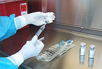
 Thin game aspiration biopsy. With this method of biopsy, a thin needle with a syringe is used. In this case, a small tumor fragment is closed. This method does not apply under suspicious mole, and it is commonly used to study lymphatic nodes located near melanoma to confirm their defeat. In some cases, when punctured lungs or liver uses computed tomography.
Surgical biopsy of lymph nodes. This method is used to remove increased lymph nodes. The procedure is usually carried out under local anesthesia. The method is used when there is a suspicion of the lesion of the lymphatic assembly, and the thin-game biopsy does not allow to identify metastases.
Marking of watchdog lymph nodes and biopsy. This method is new and promising. Watchdog lymph nodes are called because they are amazed first.
Sometimes, a small amount of radioactive substance and harmless paint is introduced to the labeling of such lymph nodes to the operation in melanoma. An hour later, the surgeon makes a small cut above the lymph node and in coloring and radioactive accumulation judges the state of the lymphatic assembly. Suspicious node is removed for microscopic research. In case of detection of metastases, the remaining lymph nodes of this area are removed. If guarding lymph nodes do not contain tumor cells, the operation to remove other lymph nodes is not shown.
In the case when the lymphatic node of large sizes is performed by a thin-game or surgical biopsy.
X-ray and other research methods
Radiography of the chest research is applied to identify melanoma metastases in the lungs.
Computed tomography (CT). With the help of X-rays, a layer-by-layer body image is obtained by rotating the apparatus around you. The method allows you to identify melanoma metastases in other organs, for example, in the liver. The method of computer tomography can be used for a sight biopsy in suspected metastase. In this case, the needle is injected into the tumor under the control of computed tomography, and the material obtained is sent to the study.
Magnetic resonance Tomography (MRI). At the same time, radio waves and strong magnets are used instead of X-rays. It is possible to obtain pictures both in transverse and longitudinal directions. Sometimes the study use contrast agents. Magnetic resonance tomography is especially useful in the study of the head and spinal cord.
Positron-emission tomography (PET). With this method, glucose is used that contains a radioactive substance. Special camera determines radioactivity. Tumor cells absorb a large number of radioactive glucose due to increased metabolism. The method is used when there is a suspicion of the distribution of the process, but not known the location of metastases. With this method, the whole body is investigated.
Scanning with radioactive materials. With this procedure, a radioactive substance is introduced into the vein in very low doses. Then the special chamber estimates radioactivity throughout the body. If melanoma spread into bones or liver, then the method will allow this to determine.