Ways of infection by fasciolesis, symptoms and diagnostics of fasciolez. Treating Fazioleza.
Content
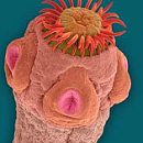
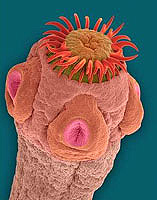
Helmintosis caused by trematodes from the family «FasciOLidae» See «Fasciola» (Common Faziol, 2-3 cm long) and «Fasciola Gigantica» (Giant Faziol, is considered the most pathogenic, reaches 3-8 cm). Female shapes parasitize in herbivores (horses, cows, sheep, pigs) and a person in the bile stocks of the liver.
Faziol ordinary is common everywhere, and a giant fasciola is found in the regions with a warm climate (in the Astrakhan and Guriev regions, Transcaucasia, the republics of Central Asia and Kazakhstan), as well as in the southern regions of Yakutia.
Sources of infection
The source of animal infection is invasive mollusks that are intermediate owners: for «F.Hepatica» - Small pondovik for «F.Gigantica» - Correspondent Prudovik. The source of invasion (infection) for humans of the liver facility of an ordinary is a small horned cattle, a giant fasciole – cows, horses, some rodents.
Ways of infection by Façiolesis
Façioli contamination occurs when drinking unsalted water, eating aquatic plants with adolescarities that attached to them, washed with dirty larvae water.
Forms of existence
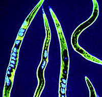 Eggs of parasites highlighted with feces fall into the water. Turn into larvae (Miracidia), which are introduced into the body of freshwater clams «Galba, Lyimnaea», Where is the most powerful reproduction of the beziol. Send from mollusk with larvae (adolescary) are attached to aquatic plants and form cysts. Plugged parasite larvae is exempt from the shell, they are introduced into the mucous membrane of the small intestine and, actively destroying the parenchyma (tissue) of the liver, penetrate into it and biliary moves, where they achieve sexual maturity.
One metatericarian (Invasive stage of larvae) It highlights up to 1000 eggs, which then fall into the intestines and are output to the external environment. In fresh water at a favorable air temperature (+ 20-30 s), Miracidium is formed from the egg, which goes into the water. Its length is 0.15mm. From the moment of entering the adolescarium (Lichwater Stage of Development) In the liver to the development of the half-armed stage passes 3-4 months. Faziols in the liver of ruminants live 8-10 years old, people can parasitize up to 15 years and more.
Symptoms of Fazioleza
The main factors are toxic-enzymatic effects of parasite larvae during migration. The incubation period is 1-8 weeks.
- In the acute stage, the fasciolesis may flow as an allergic disease with a high temperature, skin rashes appear, pulmonary syndrome (volatile infiltrates, pneumonia), and jaundice often occurs, sometimes – myocarditis; Against the background of leukocytosis rises the level of eosinophils in the blood. Acute manifestations last 2 to 6 months.
- Allergic phenomena persist in the chronic stage – Skin rash, blood eosinophilia. Disruptions of the function of the biliary system with constant or bakery pains in the right hypochondrium, nausea, decreased appetite. Periodically, the yellowness of the scler and skin can appear with an increase in the level of associated bilirubin serum and alkaline phosphatase activity, and the blood protein is reduced. When connecting a bacterial infection (staphylococcus, intestinal wand), acute bouts of the type of bile colic, fever, jaundice, hepatomegaly (liver increase) may occur. It is possible to develop purulent cholecystocholangitis (combined inflammation of the gallbladder and bile ducts), liver abscesses. In children and pregnant women, fasciolesis may be complicated by pronounced anemia.
The consequences of parasitization in the body
The outcome of the disease of the fasciolesis is associated with the biology of the development of the causative agent. Young fasciols in the small intestine traumatize the mucous membrane, are embedded in capillaries, and then through a portal vein in the liver. As a result of tissue damage when migration, the fasciol in the body occurs inflammatory processes in the blood vessels, the wall of the intestine, lymphatic nodes, peritoneum, liver and biliary strokes. Accumulating in large quantities, parasites can block the bile ducts, which causes stars bile. Facioles carry a large number of pathogenic microflora from intestines to organs and fabrics. During the migration period, part of the fasciol dies, released a significant amount of toxins that have an allergic impact on the host body.
Methods of diagnosis of fascioleza
The diagnosis is established on the basis of a clinical picture, disruption of the function of the biliary system, allergic syndrome and epidemiological history (use of water plants, greenery, vegetables, fruits, washed with water from open reservoirs in areas of propagation of fasciolesis). The diagnosis is confirmed by the detection of parasite eggs and feces.
Treatment of Fazioleza
Treatment is carried out according to the scheme as when opistorhoz. Prescribed chloxyl or biltricyde, abroad for these purposes recommended to use triclaborendazole. After the course of treatment, the choleretic agents are used for 1-2 months. Durable (at least a year) Namissarization of patients.
Prevention of fascioleza
Prevention is based on the improvement of large and small cattle by treatment from fasciolez, cleaning pastures from pollution by animal feces, pasture surveys and water reservoirs for molluscs. In areas of propagation of fascioles, it is impossible to drink water from natural sources, wash it greens, vegetables and fruits used in raw form.

