Glaucoma is a disease of the eye for which the periodic or constant increase in intraocular pressure (WGD) is typical. The reason for increasing IOP is a violation of the circulation of intraocular fluid (reduction of its outflow). Atrophy of the optic nerve occurs due to the increased WFD, which over time leads to full loss of vision.
Glaucoma is primarily threatened by those whose relatives were subject to this disease (the disease is more often transmitted through the generation: if the grandfather sick, then the grasses almost certainly develops glaucoma). Also in the risk group, people suffering from myopia, diabetes, endocrine diseases. Glaucoma may be congenital, matters and age: acquired glaucoma most often appears after 60-65 years, and in some cases after 40 years.
As glaucoma arises
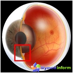
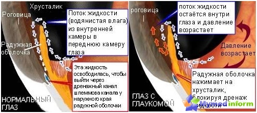
In the eye is constantly produced by a water-bearing moisture, which then goes into the back chamber - a small space between the iris and lens. Then through the pupil moisture falls into the front chamber - between the cornea and the iris. In the angle of the front chamber, where the cornea and iris are converged, the drainage eye system is. Through the drainage system of moisture comes out of the eye and enters the bloodstream. If the outflow is broken - rises in. The longer the WFD remains elevated, the stronger the visual nerve. Over time, the fibers of the optic nerve begin to die and the person will gradually blind. So that the BGD remains normal, the balance between the production of moisture and its outflow should be observed.
Normally, the eye pressure does not exceed 16-27 mm RT. Art. Although the glaucoma of the WGD can be both normal (or not much to go beyond the norm) and elevated. But if the pressure exceeds 35 mm RT. Art. and remains for a long time, must be treated.
Symptoms
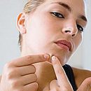
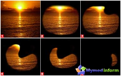
If intraocular pressure increases, a person is experiencing a number of unpleasant symptoms: vision becomes blurred, the feeling that you look «Through the grid». When looking at a lamp or any other light source before your eyes, broken rainbow circles appear. Worsening vision at dusk, there is a thread in the eyes, tear, feeling of gravity and tension, pain around the eyes.
It happens that the person does not notice the increased VGD (glaucoma may pass asymptomatic at the first stages) and appeals to the doctor only when the field of view begins to narrow. Our site draws your attention to the fact that in this case you can only stop the loss of vision, it is impossible to return the normal field of view.
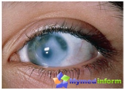
A sharp attack of glaucoma develops suddenly. There is a strong pain in the eye and the corresponding half of the head, the pain in the back of the head, nausea, vomiting, the total weakness. The symptoms of the attack are very similar to the manifestations of a hypertensive crisis or poisoning - because of this, the patient does not receive assistance in time and can lose sight.
Here are the symptoms for which you can define the attack of glaucoma: the eyelids will appear, the eyes are blushing, the cornea is cruck, the pupil is expanded and has an irregular shape. Vision is sharply reduced. If you touch your eyes with your fingers (closed), it will be very hard - due to increased VGD. To help the patient, you need to immediately dig a 2% solution of pylocarpine, take 0.25 g of diakar (diuret tool required to remove fluid from the body) and consult a doctor as soon as possible.
Diagnosis of glaucoma
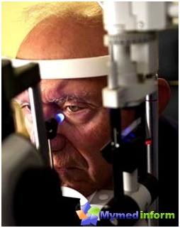
To make a diagnosis of an ophthalmologist's doctor, it is necessary to conduct a number of simple (for the patient) surveys: determination of visual acuity (visometry), measurement of intraocular pressure (tonometry), examination of the field of view (perimetry), determination of the state of the optic nerve during an eye examination (ophthalmoscopy). All the necessary devices are in any ophthalmologist. An experienced doctor can diagnose glaucoma even after palpation of eyeballs and ophthalmoscopy.
Glaucoma classification
As already mentioned, glaucoma can be both congenital and acquired during life.
Congenital can manifest itself both in childhood (up to 10 years old, most cases) and at a later age.
Acquired glaucoma, depending on the cause of its development, is divided into primary and secondary. The primary glaucoma is developing due to the disturbance of the outflow of water-melting moisture from the eye and, as a result, increasing the VGD. Secondary glaucomas include all those that have appeared as a complication of other diseases (diabetes, cataracts, thrombosis), injury and inflammation of the eye and other organs.
According to the mechanism of increasing VGD - the acquired glaucoma is divided into an open-dectorial and closed.
With an open-angle glaucoma, the drainage system of the eye gradually changes, which flows periodically the pressure and suffers from the visual nerve. With a closed-coronal glaucoma, the outflow of intraocular fluid overlaps the root of the iris, which closes the angle of the front chamber of the eye.
Depending on how badly the optic nerve glaucoma is divided into 4 stages. At the 1st stage (initial), the field of view has not yet been narrowed, but when an eye-examination of the eye can be seen. For the 2nd stage (developed glaucoma), the narrowing of the field of view is already characterized, and the changes in the eye day are pronounced stronger. At the 3rd stage (far-seated glaucoma), the field of view is narrowed concentrically, and on the 4th (terminal glaucoma) patient completely loses his eyesight or only light supply remains.
Treatment of glaucoma

In the treatment of glaucoma, there are three main directions: therapeutic, laser and surgical.
In the therapeutic treatment, the patient is prescribed drugs lowering intraocular pressure, and in addition to them - drugs that improve blood supply and normalizing metabolism in eye tissues.
Medicines that reduce IGDs should be regularly Reduce pressure, But it happens that the patient has resistant to the action of droplets - and then you have to select another drug. In the first weeks of appointment of anti-flame droplets, the patient must regularly visit the eyepiece to keep control over the state of intraocular pressure. If the drops have a positive effect, the patient will need to visit the doctor only once every 2-3 months. In some cases, the drops are so successful that with their help it is possible to control WGD for several years - you only need to change the drug only once a year, so that there is no addiction.
To lower the eye pressure, use two types of droplets: drops that improve the outflow of intraocular fluid (pylocarpine, isocarpine, etc.) and drops that reduce the production of intraocular fluid (clonidine, beta, sandy). Our site strongly recommends: Never mix drops. This wild «cocktail» From different drugs will not bring any benefit of the eyes, on the contrary, drops will lose their healing properties.
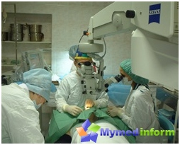
If drops do not give effect, you have to make an operation. Most often, first carry out a laser operation. The most effective are laser iridotomy (iridectomy) and laser trabewallasty.
The essence of laser iridectomy is that the doctor using a laser beam makes a small hole in the iris. Through this hole, the intraocular fluid begins to leave the eye without obstacles, the VGD is normalized. This operation resort to the treatment of closed-curved glaucoma.
Laser trabewallasty is carried out with an open-angle glaucoma. It also contributes to the normalization of the WGD, but its principle is a bit different: a laser beam causes several microscopically small burns on the iris, which increases the permeability of fluid iris.
Advantages of laser operations:
- Outflow of intraocular fluid occurs in natural ways;
- For the operation, it is sufficient to drip anesthetic to the eye;
- The operation can be carried out in an outpatient basis;
- The term of rehabilitation is minimal.
Disadvantages of laser operations:
- DISTRIBUTY OF THE EFFECT OF OPERATION (1-2 years);
- During the operation, damage to the epithelium of the cornea, crystal capsules and iris vessels are possible;
- The reaction to the laser operation may be increased in the first hours after the operation;
- After surgery, fabrics can form in places of exposure to laser.
The decision on the need for a surgical operation is made in each individual case on the basis of the type of glaucoma, the testimony of intraocular pressure and the level of fluid outflow from the eye, field of view, as well as a general patient health assessment. The most common types of surgical surgery during glaucoma are trabcularctomy and impermeal deep sclerctomy (NGSE).
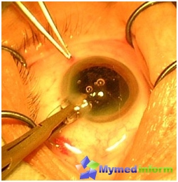
In Trabeculectomy, new ways of outflow of intraocular fluid are created. During operation, the doctor removes the fabric site and creates a direct message between the front chamber of the eye and the subcactive space. This operation is inherently relatively lightweight, and brings quite good results. But not rare the results of trabeculectomy not only are not preserved for a long time, but, on the contrary, a number of negative consequences appear. On the site of operational intervention there are scars, t.E. re-blocks fluid outflow. And what is characteristic, the re-operation will no longer have the proper effect. In some cases, on the contrary, the outflow of intraocular fluid begins to go much faster than it reproduces - it will also negatively affect the eye.
The essence of NGSE is the surgical thinning of the peripheral sections of the cornea. Through these thinned moisture places, freely circulates and easily leaves the eye. After NGSE, the patient may return to the usual way of life after 2-3 days. But that the eye pressure remains stable, periodic (2 times a year) Supporting therapy courses are required: drugs that improve the blood supply of the eyeball and optic nerve (Cavinton, Picikalon, Mildronat), physiotocreders.
As you can see, glaucoma is a pretty serious illness. Therefore, if you feel that vision is cloudy or even just light pain in the eyes, do not be lazy to go to the doctor. As prevention, after 40 years, it is desirable to periodically visit the oculist and without visible. Those violations coming together with the development of glaucoma are no longer restored - you must remember and take care of the state of your eyes.