What is hydronephrosis? Why hydronephrosis occurs? As manifests and treated hydronephrosis? Read in this article.
Content
Hydronephrosis or obstruction of the gluhan and ureteral segment
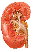
 Hydronephrosis is an expansion of a collective kidney system (especially pelvic), resulting from the presence of an obstacle to the urine output at the junction area of the locher and the ureter (in the area of the piiveureteral segment).
Urinary Ways include (from top to bottom) renal cups, kidney pelvis, ureters, bladder, urethra. Lohanks and cups together make up a collective kidney system.
The pronounced obstacles to the outflow of urine from the kidneys lead to a significant expansion of the locher and, often, to an irreversible impaired of the kidney function. The degree of expansion of the collective kidney system is proportional to the pressure of urine in it and varies widely.
A small obstacle to the output of urine, causes moderate expansion of the pellektasia and is usually not accompanied by a disruption of renal functions, but only increases the risk of pyelonephritis.
Causes of hydronephrosis in children
In children in the overwhelming majority of cases, congenital hydronephrosis of the ICD, due to anatomical causes. There are external and internal causes of hydronephrosis. Internal reason – Congenital narrowing of the ureter due to the underdevelopment of his lumen, occurs more often than others. External reasons – Abnormal extinguishing of the ureter from the pelvis and an additional vessel causing the ureter compression.
Manifestations, symptoms of hydronephrosis
Hydronephrosis is included in a group of diseases accompanied by the expansion of the renal lochank (pyelectasia), which is easy to detect when the fetus ultrasound. Therefore, most of the hydronephrosis is detected by intrauterine. If the diagnosis was not installed before the birth of a child, hydronephrosis can appear in the blood of blood in the urine (hematuria), infection of the urinary system, pain in the abdomen, or when the volumetric education in the abdominal cavity is detected.
Studies at hydronephrosis
The first step towards the diagnosis of hydronephrosis is an ultrasound of the fetus. The collective kidney system is visible in ultrasound examination from 15 weeks of the intrauterine period. The first sign when ultrasound is the expansion of the jelly. If after the birth of the child, the expansion of the pelvis is preserved, then a children's urologist solves the question of the need for a more in-depth urological examination. In suspected, the presence of hydronephrosis, the child must pass the following surveys:
Ultrasound kidneys and bladder before and after urination. Specialist in ultrasound can see signs of damage to the kidney parenchyma, distinguish a weak, average and pronounced degrees of hydronephrosis. In questionable results, ultrasound with water load and diuretics can be performed, which allows you to more accurately assess the degree of obstruction of the gluing ureteral segment.
Miking cysturketography – X-ray-contrast examination of the bladder and urethra is performed in suspected bubble-ureter reflux or hindered urine outflow from the bladder.
Excretory (intravenous) urography – After intravenous administration, the X-ray contrast is derived by the kidneys, and their collective systems become visible on X-rays. Research allows us to estimate the degree of obstruction.
NEFROMYCINTIGRAGY – Radioisotope examination of the kidneys. Used to estimate the kidney function and the degree of violation of urine outflow.
Based on the studies, the specialist should decide how serious the obstruction of the gluing ureteral segment is, whether it is a threat to the kidney or can be resolved independently. Newborn diagnosis becomes obvious often only after 3-4 weeks after birth. During the first 3 weeks after birth, the water exchange in the body of the newborn and the kidney function varies considerably, and with them the sizes of loyal.
Treatment of hydronephrosis
Initial manifestations of hydronephrosis often disappear independently, but sometimes progress. The observation of a specialist with the implementation of the ultrasound is shown 2-4 times a year, in the first 3 years of life, and once a year at older age.
The average degree of hydronephrosis, starting with hydronephrosis of 2 degrees, may have, both positive and negative dynamics. With an increase in the expansion of the jack in the process of observation, it is necessary to conduct operational treatment. Ultrasound in the first year of life at the average degree of hydronephrosis are held every 2-3 months.
Pronounced hydronephrosis with a sharp impaired of urine outflow from the kidney requires the execution of a surgical operation without delay.
Operation in hydronephrosis
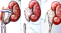
 Operation in hydronephrosis lies in the excision of a narrow portion of the ureter and the formation of a new wide compound (anastomosis, cooler) between the ureter and the kidney laughter. Called operation – Peloplastic.
The most common technique of surgery - Heins-Andersena pyeloplasty. The narrowed ureteral place is usually located directly near the renal loin. After cutting off the ureter, its closest to the kidney area dishess longitudinally, after which the edges of the ureter cut are stitched with the edges of a symmetric (congruent) longitudinal cut on the lochank. Usually, after the operation, leave a tube conducted through the place of the connection of the ureter and pelvis, to ensure the uniform lumen of the coherence and avoid its sticking and deformation. The second end of the tube can be removed in the bladder (stent inner drainage) or through the kidney tissue (catheter-dwarf).
Duration of finding a child in the hospital after surgery
The duration of the child's stay in the hospital after the operation depends on the method of urine leads from the operated kidney. When installing a stent inner drainage, additional drainage for urine leads from the kidneys is not required, and the stationary postoperative period is reduced to 5-9 days. The stent is removed in a month – A half after surgery through a thin tool entered by urethra.
If it is not installed during the operation, a stent, but a catheter, which is derived out through the kidney, is established in parallel, the Drainage tube (nephrosta) is installed to provide free urine outflow from the operated kidney. In this case, more prolonged finding a child in the hospital - about 3 weeks. The choice of the urine leads is made by the surgeon during the operation.
Features of treatment and anesthesia are determined taking into account discussed with the operating surgeon
The effectiveness of pyeloplasty
The effectiveness of pyeloplasty is about 92-95%. After performing the operation, the kidney function is almost always improved and in a number of observations reaches the function of a healthy kidney. At the same time, structural changes in kidney (deformation of cups, a reduction in the thickness of the parenchyma) can persist. Especially significant residual changes are observed with a pronounced hydronephrosis.
Forecasting the course of hydronephrosis in a newborn
Method allowing to determine how hydronephrosis will develop in the newborn, currently does not exist. Therefore, the most correct approach is to observe the condition of the kidney in the dynamics of an experienced specialist urologist. The main estimate method for dynamic observation is ultrasound. The complexity of predicting the development of hydronephrosis in a newborn is determined by an unstable water exchange, changing the function of the kidney, as well as the possibility of ripening (maturation) of its organs and tissues. These processes can lead to the disappearance of the expansion of the locher or stabilize its size. At the same time, with long intervals between the inspections (more than 2 months), you can skip the worsening of the kidney state and be late with the operation.


