What are the types of hemangiom? Where hemangi is a favorite place? Read in this article.
Content
Types of hemangiom
Distinguish simple hemangioma taking place from the smallest blood vessels – capillaries; Cavernous hemangiomas consisting of cavities filled with blood; Combined hemangiomes having subcutaneous cavernous and skin capillary parts; and mixed, consisting not only of blood vessels, but also include other types of fabrics.
Simple hemangioma
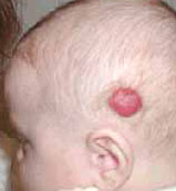
Simple hemangioma have a red or blue-bug color, are located superficially, clearly excluded, affect the skin and several millimeters of the subcutaneous fat layer, grow mainly on the parties.
Surface hemangiom smooth, less often – uneven, sometimes somewhat protruding above the skin. When pressing hemangioma is pale, but then restore your color again.
Cavernous hemangiomas
Cavernous hemangiomas are located under the skin in the form of a limited nodular formation, soft-elastic consistency and consist of a different amount of cavities - a cavern filled with blood. They look like a tumor-like formation, emanating from the subcutaneous fat layer, in the indoor unchanged or cyanotic on the top of the skin, with the growth of vascular tumor, the skin acquires a synebaggrot color.
When pressing hemangioma, it falls on and pale (due to blood outflow), when crying, crying and kashek, the child increases and strains (an erectile symptom resulting from blood inflow). In hemangiomas, the skin is usually clearly detected by the symptom of temperature asymmetry – Vascular tumor on the touch of hotly surrounding healthy fabrics.
Combined hemangiomas
Combined hemangiomas are a combination of surface and subcutaneous hemangiom (simple and cavernous). They manifest themselves clinically depending on the combination and predominance of one or another part of the vascular tumor.
Mixed hemangiomas
Mixed hemangioma consist of tumor cells emanating from vessels and other tissues (angiofibrom, hemlimphangioma, angioedema, etc.). Their appearance, color and consistency are determined by the composition of the vascular tumor tissues.
Hemangioma may be single and multiple. Most of the hemangiomas of large sizes were single, and multiple tumors, as a rule, are small. A group of patients with small and medium hemangiomas surpasses a group with large and extensive hemangiomas, the treatment of which represents the greatest difficulties. The choice of the treatment method of extensive hemangiomes is very problematic.
Location hemangiom
 The favorite localization of hemangiom is the area of the head and neck. With this location, the surgeon, assigning treatment, seeks to get not only the healing effect, but the most perfect cosmetic and functional result. In the localizations of hemangiom on the body and limbs, the cosmetic result of treatment has much less. When localizing hemangiom in the body of the body, the largest interest in practical doctors is their location in the chest and crotch area. Although the localization of the chest boys and girls is the same, the approach to the treatment of these hemangioms has significant differences. A feature of the course of hemangiom in the crotch and external genital organs is their tendency to frequent ulceration.
Children with hemangiomas can develop thrombocytopenia – Reducing blood cells responsible for stopping bleeding due to cluster and thrombocyte death in vascular tumor (Casabach syndrome – Merritt). As noted above, the peculiarity of the flow of some hemangiom is their tendency to frequent ulceration and self-esteem, which causes doctors with special care to treat or, if possible, avoid it. At the location of the ulcendous vascular tumor in the area of the lips, the act of sucking was disturbed, in the nose area, especially when the tumor filled a completely nasal stroke, – Act of breathing. The ulceration of hemangioma in the field of external genital organs, especially in girls, causes severe pain and burning sensation, which is hardly reflected in general condition.


