Deforming arthrosis - a disease characterized by chronic, continuously progressive current. Diagnosis of osteoarthrosis, stage of illness and their x-ray signs. The difference between arthrosis 1 degree from arthrosis 2 and 3 degrees.
Content
 Osteoarthrosis — Degenerative — Dystrophic disease arising from damage to cartilage covering the articular surfaces of the epiphysis of bones forming the articulation. In the pathological process, not only cartilage is gradually involved, but all the tissues of the joint, including the synovial shell, subchondral bone, the articular capsule, ligaments, holding the joint and muscles leading it in motion. The defeat of one joint leads to an increase in the load on others, therefore, creates conditions for the development of arthrosis of another localization.
The diagnosis of deforming osteoarthrosis includes:
- analysis of clinical symptoms;
- General and biochemical blood tests;
- radiography;
- Computer tomography, magnetic resonance tomography;
- Sonography (ultrasound joint);
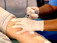
- Arthroscopy, biopsy and biochemical study of synovial fluid.
If necessary, the examination can be complemented by other methods.
Clinical diagnostics of arthrosis, its stage
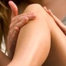
 Arthrosis of 1 degree can proceed asymptomaticly, only some increased fatigue of the limb is possible. However, with a detailed examination, with arthroscopy and biochemical study of intra-articular fluid, signs of metabolic disorders in the tissues of the joint can be detected at this stage of the disease. These violations are caused by dystrophy of cartilage and lead to the loss of their abilities to withstand ordinary loads.
Arthrosis of 2 degrees characterized by the destruction of the articular cartilage and the formation of osteophytes, has pronounced clinical symptoms. Typically appearance «starting pain» and stiffness. Pain in the joint is intensified by the end of the day, but passes after a long rest. The manifestation of arthrosis of the 2 degree is a crunch in the joints, their initial deformation and thickening due to the growth of regional osteophytes. Involvement in the process of a ligament apparatus on the one hand leads to the pathological mobility of the joint, on the other — leads to limitation of the volume of movements.
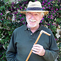
The sign of osteoarthrosis of the third stage, in contrast to the arthrosis 1 2, is the pronounced joint deformation and the change in the axis of the limb. Causes are crumbling in distortion of the form of the joint area of the joint as a result of the destruction of cartilage and degenerative changes in the bones forming the articulation. Restricting the mobility of the joint reaches the maximum. There is a contracture and ankylosis that deprive the patient the possibility of independent movement without the use of crutches or canes.
X-ray diagnosis of osteoarthritis
 The main criteria for which osteoarthritis is diagnosed, the stage of its development and degree — These are radiographic signs of degenerative-dystrophic and structural changes in the joint.
X-ray symptoms osteoarthrosis
- The narrowing of the articular gap — Certificate of significant changes in the articular cartilage and weakening of the ligament apparatus holding bone articulation.
- Osteophytes arise as a result of proliferation of edge departments of the articular cartilage, as the reaction of the subchondral plate of the bone to increase the load. The first osteophytes appear as small bone cloves along the edges of the articular surfaces, then they grow up, turn into lips and spikes deforming the joint.
- Changes in bone epiphysis tissue begin with sclerosis, incurring blood vessels and bone marrow replacement with a connecting tissue, noticeable on a radiograph as a zone of enlightenment. Blood impairment in this area is systematically leading to bone tissue atrophy and appearing in it, in the subchondral zone, cyst looking in the picture as rounded black education.
- The unstability of the joint is manifested by sublifiers, change in the axis of the limb.
The deforming arthrosis of the first degree on the radiograph looks like a slight narrowing of the articular gap and a slight seal (sclerosis) of the bone in the roasting zone. Changes are still insignificant and additional research is required for more accurate diagnostics.
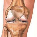
Changes in the joint depends on the level of load on it. The deforming arthrosis of the knee joint 1 in this stage is characterized by the development of compensatory and adaptive processes in the bone and regional bone growths (osteophytes).
X-ray signs of the second degree — The pronounced narrowing of the articular gap, osteophytes, subchondral osteosclerosis.
That arthrosis «increased» Of the 2 degrees and reached the third, the almost complete absence of the articular slit, a distinct subchondral sclerosis and the appearance of cystic formations in the epiphyses of bones, the deformation of the joint and distortion of the normal axis of the limb.