Material ultrasound: Normal echocartine, decoding and research results.
Content
Ultrasound of the breast
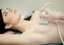
Ultrasound of the mammary glands applied by mammologists, oncologists for diagnosis. The study does not require special training, it is carried out quickly and painlessly.
X-ray mammography has greater accuracy and informative compared to ultrasound. Optimal Days for Procedure, — From the sixth to the tenth day from the start of menstruation.
During the ultrasound of the mammary glands, they define:
- Structure. Uniformity structure — Sign of healthy breast. Availability of sections with increased or reduced echogenicity — Sign of focal or diffuse changes;
- Ehogenesis — The degree of reflection of sound waves. The higher the indicator, the higher the tissue density of the breast. The minimum density has cysts containing liquid (rounded black formations, possibly with a liquid level or inhomogeneous content). Maximum calcinates (bright white, with clear contours, acoustic shadow);
- Type of tumor. Benign formations usually have increased echogenicity, clear contours, a homogeneous structure. Malignant — inhomogeneous structure, blurred contours, possibly with reduced echogenicity;
- Retromammar space, regional lymph nodes, other anatomical education and their characteristics.
Ultrasound of the mammary glands in young women and women with fibrous-cystic mastopathy can be applied on a par with mammography.