Often you have to hear: «I had a doctor, appointed CT or MRI for examination, something like that, I don't remember exactly, and what's the difference?» And really, what's the difference? What survey should choose which one is informative?
Content
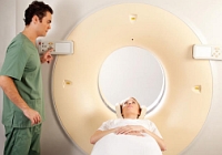 Today it is difficult to imagine the development of medicine without computer or magnetic resonance tomography. How often, in the diagnosis of certain diseases, these magical letters sound — CT and MRI. What do they mean?
In the title of both diagnostic methods, the word is used «tomography», that with Greek translates as «Study cut». Both of these methods — non-invasive methods of diagnosing diseases, that is, carried out without penetration into the inner media. However, CT and MRI are based on various physico-chemical phenomena.
Computer and magnetic resonance tomography reflect the achievement of ultrahigh medical technologies. During these diagnostic procedures, an image is displayed on the monitor screen, which is obtained when carrying out layer-by-layer sections of organs and tissues. Tomograph sends electronic impulses, and the capturing equipment reacts to the effect occurring in the body and transforms it into a three-dimensional image. Each layered slice after scanning is being analyzed and interpretation, which allows the doctor to make certain conclusions.
Principle of CT operation
Computed tomography is very similar to the traditional radiography, although it is used, a fairly simple principle. An x-ray beam affects the research area. Passing through the fabrics with different density, rays are absorbed, there is a layered image of organs and tissues. The computer processes the data obtained, which allows you to see the features of the body under study.
When analyzing tomograms, a specialist has the ability to determine the exact location and size of the organ, its anatomy and the presence of pathology. It should be remembered that during the CT, the radiation load is increased, it means that it should be refrained from the multiple application of this diagnostic method.
In the case of the use of CT, a specialist can not only see fabrics, but also to investigate their X-ray density, which changes in the presence of certain diseases. CT preferable for diseases of the base of the skull, chest, pelvis, abdomen, is used to diagnose morphological changes in internal organs, detecting tumors, internal bleeding. The reliability of the method ranges from 90 to 100%.
CT is the most informative in the following cases:
- brain injuries and skull bones;
- brain tumors, brainwater disorders;
- damage to bones, the base of the skull, the incomplete sinuses, the temporal bones;
- aneurysms and diseases of vessels;
- diseases of the spine, osteoporosis, intervertebral hernia, scoliosis;
- Pathology of chest and mediastinum;
- suspected lung cancer;
- Damage and diseases of bones.
MRI features
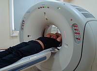 One of the safest research methods, but more expensive — magnetic resonance therapy when the image is evaluated visually. If computed tomography is based on X-ray rays, which give an idea of the physical condition of the substance, then during the study with MRI, constant and pulsating magnetic fields are used, as well as radio frequency radiation, which helps to assess the chemical structure of tissues in the body.
During the procedure, the patient is placed in the region of the magnetic field. At the same time, the device allocates a radio frequency pulse, human tissue molecules enter the resonance with him, and the name of this method occurred from here: magnetic resonance tomography. MRI — a rather long procedure, while absolutely safe and painless, which allows you to assign it to children and pregnant women.
This diagnostic method gives more information with diffuse and focal lesions of the structures of the brain, spinal cord pathology, damage to cartilage tissues, the study of the spine, joints, liver, spleen, pancreas, kidneys and sexual system. In some cases, when conducting differential diagnosis in medical practice, it is resorted to the joint implementation of MRI and CT.
The maximum information content of MRI is noted during the diagnosis:
- brain tumors, pituitary glands and intracranial nerves;
- spinal cord diseases;
- peeling syndrome in the spine;
- diseases of the joints and joint surfaces, a binder apparatus and muscle tissue.
MRI with contrasting is widely used in oncological practice to determine the degree of propagation of the tumor process, the stages of metastasis and the stage of the disease.
Precautions for CT and MRI
MRI can not be carried out to patients with inadequate behavior and in violation of life functions requiring hardware correction.
The patient is obliged to warn a doctor about the presence of a foreign metal body in the body, such as metal fragments, an artificial cardiac rhythm driver, metal implants, non-removable dentures, surgical brackets, and T. D.
The contraindication to the MRI is overweight and claustrophobia, that is, fear of indoors.
Due to the availability of radiation load CT, it is not recommended to carry out pregnant women.


