From the conversation of two young girlfriends: «You imagine yesterday I was on the ultrasound, and the doctor said that I had a girl, and even gave me to see her. What is her pretty!»
Content
- What is ultrasound?
- The most common ultrasound studies
- Ultrasound during pregnancy: your baby's photo, now color!
 20 more–30 years ago three mysterious letters — «Ultrasound» — little about what they also spoke and ordinary patient, and even some doctors. And today, without this examination, it is difficult to present medicine, especially some of its industry. It is hardly possible to find an adult person who does not know what an ultrasound is, or who at least once in life did not pass this type of diagnosis.
Many experts argue that the opening of an ultrasonic wave has become the greatest breakthrough in medicine. It allowed the doctors to evaluate the structure of almost all organs, identify various diseases, differentiate pathological processes and immediately proceed to treatment, sometimes saving the patient from the operation and still invisible complications.
What is ultrasound?
In general terms, we all imagine what it is and what is the value of ultrasound. The fact that this is a modern method becomes clear as soon as you crossed the threshold of the Cabinet ultrasound. The Cabinet Equipment itself transfers the patient into the situation of high technologies that have become available to a person only in recent years. The use of an ultrasound scanner allows a specialist to perform research and give an accurate conclusion about the state of health.
The ultrasound method is very in demand today, and there is one explanations and reasons. The main advantages of this study are several.
- First, full safety and lack of ionizing radiation. This feature should be attributed to one of the main advantages of this method, which is based on a piezoelectric effect.
- Secondly, non-invasiveness. Ultrasound is carried out without disrupting the integrity of the skin and the introduction of toxic contrast agents.
- Third, painlessness, simplicity and speed of procedures.
- Fourth, high informativeness, which seems incredible when the asymptomatic disease occurs to be diagnosed at the initial stage.
With confidence it can be said that the scope of ultrasound research does not have contraindications and does not know the boundaries. With the help of ultrasound, almost any organ of the human body can be explored.
This may be, for example, the study of the abdominal bodies, thyroid glands, a small pelvic organs and a reproductive system of a man or a woman. With the help of ultrasound waves, women are conducted by dairy, and men — prostatic gland. Of great importance is ultrasound when examining pregnant women and fetus. «Photo for mom and dad» It became a traditional souvenir from the Uzi Cabinet during prenatal diagnostics.
Ultrasound examination of the heart, liver or pancreas can become «Rescue circle» For those who are sinking in the depths of an unidentified diagnosis. At the initial stage of the disease or with its asymptomatic flow, it plays a huge role.
The most common ultrasound studies
 Take for example several organs of the human body and consider the possibilities of ultrasound.
For example, heart. In what cases is the ultrasound of the heart or echocardiography? The study is carried out in cardiac pains, shortness, rapid heartbeat of incomprehensible origin, interruptions in the work of the heart, improving blood pressure and with suspected heart defects.
Ultrasound examination of the vascular of the lower extremities is shown in pain and seizures of legs, edema, their inexplicable increased fatigue. Inspection allows the doctor to establish the degree of varicose disease, the permeability of the main vessels, and also give recommendations about the methods of treatment of vessels.
Diagnosis of diseases of the abdominal organs allows you to clearly set the size and structure of internal organs, for example, liver, spleen, pancreas, and diagnose the presence of stones in a busty bubble or kidneys. The method makes it possible to recognize various neoplasms and changing the structure of lymphatic nodes.
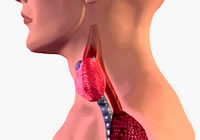
Ultrasonic study of the thyroid gland helps to estimate the structure of the organ, the presence of nodes or cystic cavities in it.
Ultrasound during pregnancy: your baby's photo, now color!
Separate topic for conversation — Ultrasound examination during pregnancy. Consider the importance and significance of this diagnostic procedure on the example of the most important event in the life of a small man and his parents. In the event of a delay of menstruation, only an ultrasound can give an answer to the main questions, whether in the uterine cavity pregnancy and how it develops.
Of course, a young mother sees in the procedure first of all the opportunity to get accurate information about who should be born in the family. It is clear that this is an important point, but not the main thing, because the doctor when conducting a study first tries to understand whether there is no problems with the future child, whether the rates of its growth correspond to certain standards.
The first planned ultrasound (or ultrasonic screening of the first trimester) is carried out on the period of pregnancy 11–12 weeks. The doctor determines the size of the embryo, fixes the cardiac tones of the future infant, measures the size of the collar zone, assesses the anatomical features of the fetus. Specialist's eye available signs of threats of miscarriage, multiple pregnancy, as well as pathology of small pelvis and uterine organs.
The second planned ultrasound of a pregnant woman passes on a period of 18 to 21 weeks to identify the malformations of the fetus. The third (last mandatory) ultrasound examination is performed on time 32–34 weeks. At this stage, the development of the fetus is estimated, its condition before childbirth, the location of the placenta and cord and blood circulation in the system «mother — placenta — fetus», which is very important in the subsequent maintenance of pregnancy and childbirth.