In which cases is diagnosed «Irit», and in what you need to talk about iridocyclite? What are the signs of Irita and Iridochiklit? You can find out about it by reading this article.
Content
Irit and Iridocyclite: Causes and Signs
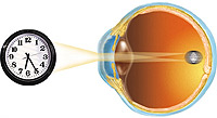
 Iridooclite - inflammation of the iris and ciliary body. There occurs on the basis of common diseases of the body (tuberculosis, rheumatism, angina, flu, polyarthritis, return typhus, gout, diabetes, syphilis, gonorrhea, etc.), diseases of teeth, the apparent sinuses of the nose, when droves with the blood flow of pathogens or their toxins in the iris and ciliary body.
Inflammation can begin in the iris (Irit), then spread to the ciliary body (cyclit) or the process simultaneously covers both shells - iridocyclite develops.
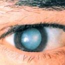
Irit is accompanied by light, tear, pain in the eye, hyperemia conjunctiva of the eyeball.
The color and drawing of the rainbow shell is changed, the pupil is narrowed, the moisture of the front chamber of the eye often becomes muddy, the spikes between the pupil edge of the iris and the leather bag appear (they are well detected when the pupil is extended).
In case of iridocyclite, the signs of cyclita join the phenomena of Irita: pain in the eye are enhanced, the acuity of sight is reduced, gray-like point deposits appear on the back surface of the horny shell, the vitreous body is observed.
Intraocular pressure is usually normal or slightly lowered. When palpation of the eyeball, an acute pain feels.
Iridocyclitis, with appropriate treatment, usually ends with recovery with recovery. With some form of iridocyclitis, for example, with rheumatic iridocyclite, there may be relapses of inflammation, which often lead to a resistant reduction in view.
With severe current iridocyclitis, complications may develop in the form of an infection of the pupil, the increments of the pupil edge to the lens bag, as a result of which the secondary glaucoma occurs. This complication threatens blindness. That is why in this case it is very important not to miss the moment and spend timely operational intervention.
Iridocyclite is detected when examining the eye with a slit lamp. In addition to the signs given above, the doctor discovers inflammation cells in the front chamber of the eye as a light suspension. With a large number of cells, they settled at the bottom of the front chamber in the form of a drop of pus. Spotted protein deposits are found on the back surface of the cornea.
In addition, the doctor will surely check the rear departments of the eye to eliminate their involvement in the inflammatory process.
For the treatment of iridocyclite, anti-inflammatory preparations of steroid and nonsteroidal series are prescribed both locally in drops and ointments and in injections and tablets. In addition, drops that expand the pupil are applied to avoid soldering the pupil with a lens, narrow the extended vessels, reduce inflammation.