No one, even newborns, is not insured against retinal detachment, but with age, especially closer to 60, the risk increases significantly. The risk group also includes patients who have undergone an operation to remove cataracts that have had serious damage or inflammation of the eye, adverse heredity regarding the retinal detachment, as well as suffering from myopia. What is retinal detachment?
Content
What detached in the eyeball?
Retinal detachment is the process of separating the mesh shell eye from the vascular shell. In a healthy eye, they are closely touching. The retinal detachment often leads to a significant reduction in vision and blindness. Most often it occurs in injuries and myopia, as well as in diabetic retinopathy, intraocular tumors, dystrophy of the mesh shell and T.D.
What is retinal detachment
 Retinal detachment is the process of separating the mesh shell eye from the vascular shell. In a healthy eye, they are closely touching. The retinal detachment often leads to a significant reduction in vision and blindness. Most often it occurs in injuries and myopia, as well as in diabetic retinopathy, intraocular tumors, dystrophy of the mesh shell and T.D.
Often, our eyes are compared with the camera, in which the role of the lens is performed by the cornea and lens: they skip and refract the rays of light falling into the eye. The role of the photosensitive film is removed with a mesh shell: it displays all that we see. Retina eye thin, tender sheath similar to light veil. It consists of many nerve cells and their processes. Cells that perceive light are called molds and chopsticks. In the retina, the eye of the man's chopsticks is many times more than the columns: the sticks of about 170 million, the columns of 8 million. Under the action of light in these cells, the visual pigment disintegrates and special substances are formed. They cause a kind of electric discharge, which, as for electrical wires, is transmitted by retinal nervous cells, and then by visual nerve in the brain. In the cerebral cortex, or rather, in its occipital part, this electrical signal turns into a visual image.
Sticks and columns are distributed in the retina unevenly. Each type of these nerve cells has its own singular functions. Columns are concentrated in the center of the retina, in the place where the rays fall from the objects under consideration. They provide a person with high visual sharpness, perception of all colors and their shades. Wands provide us with twilight and lateral vision, that is, allow you to see at dusk and navigate in space. The sticks are relatively evenly allocated throughout the retina, but their number increases as removal from the center. Norma retina closely adjacent to the vascular shell from which it gets food. Why is the retina, so firmly placed in the eyeball, still moves from his place, peels off, ceasing to function. What are the reasons for this?
Causes of retinal detachment
 Experts allocate the main cause of this mesh shell gaps. The retina is not shifted from its place if it is sealed (retains its integrity) and there is no gap in it. If the gap formed, then through it the vitreous body fluid penetrates under the retina and flaps it from the vascular sheath.
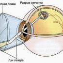
The main reason for the formation of the retinal ripping of the vitreous body when changing its normal state. This process occurs in this way: Normally, the vitreous body resembles a transparent jelly. In some eye diseases it changes, becomes muddy with dense heavy, t.E. compacted fibers that are associated with the retina. When driving, the eye is pulled by the retina, which can lead to her rupture.
Of course, no one, even newborns, are not insured against retinal detachment, but with age, especially closer to 60, the risk increases significantly. The risk group also includes patients who have undergone an operation to remove cataracts that have serious damage or inflammation of the eye, unfavorable heredity regarding the retinal detachment, as well as suffering from myopia (factor that increases the likelihood of retinal detachment, because at myopia the eyeball stretches and presses on the retina.) So, for example, about 20 thousand every year. Americans detect retinal detachment.
The retinal detachment occurs when the gel of the vitreous body during deformation presses with the power on the retina and breaks it, forming a hole. If the retina is damaged, the liquid from the vitreous body can be found through the hole and move the retina from the supply of its vascular eye sheath.
Rales of retina can also occur with its dystrophy (thinning). Large breaks often occur during eye injury.
Symptoms of retinal detachment
The success of the treatment of retinal detractor depends on timely appeal to the doctor. The earlier the disease will be detected and its reasons are found, the faster it is necessary to carry out the necessary treatment and the better the result will be.
Sometimes the appearance of floating points or sparks in sight is a sign of retinal detachment. This complication occurs when the inner layers of the retina - a thin photosensitive nervous tissue, lining the inner surface of the eye and conducting electrical impulses from the optic nerve to the brain, are separated from its pigment layer.
What can indicate the beginning of the retinal detachment?
- appearance of pellets in front of the eye. Patients are unsuccessfully trying to eliminate her independently, washing the eyes of tea or drowning drops. In this case, it is important to remember and tell a doctor, which side originally appeared, since it may increase and take the whole field of view
- Flashing in the form of sparks and lightning are also a characteristic feature of what is happening retinal detachment.
- Distortion of the letters under consideration, items, falling out of the field of view of their individual sites indicates that the detachment captured the retinal center.
- Sometimes patients note that after sleeping the vision is somewhat improved. This is explained by the fact that with the horizontal position of the body, the retina returns to his place, and when a person takes a vertical position, it again moves away from the vascular shell and impacts are renewed.
The retinal detachment cannot be cured by any drops, tablets or injections. The only way to restore vision and save the eye urgent operation.
With retinal detachment, nervous cells, sticks and columns die, and the longer the detachment there is, the more these cells die and the worse the restoration of vision even after a successful operation.
Remember! If you run a disease, there may be a danger of chronic inflammation, the development of cataracts, the eye can completely lose the ability to see. Especially vigilant need to be that people who have already had a retinal detachment on one eye. They should be periodically examined from an ophthalmologist. And when suspicious symptoms appear, immediately contact your doctor.