Increased intracranial pressure has a negative impact on the work of the central nervous system, which leads to the appearance of characteristic symptoms of the disease, primarily headaches. The gold standard of diagnostics are tomography of the brain and the eye picture.
Content
The main symptoms of intracranial hypertension
Increased pressure on the brain substance capable of disrupting the work of the central nervous system. It is when violating the work of the central nervous system and the characteristic symptoms of the disease appear:
- severity in the head or headaches growing in the morning or in the second half of the night
- In severe cases, nausea and / or vomiting in the morning
- Vegeth vascular dystonia (sweating, fall or enhancement of blood pressure, heartbeat, pre-real states, etc.)
- Tired, «Recess», Easy deduction with loads for work or school
- nervousness
- «Synyaki» under the gases (if you pull the skin under the eyes in the area «Sinyaka» Enhanced small veins are visible)
- It is possible to reduce sexual attraction, potency
If the human body is in a horizontal position, the liquor is more active, and is slower, so intracranial pressure and its symptoms have a property to reach a peak in the second half of the night or by morning. Intracranial pressure is higher than the lower pressure atmospheric, so the deterioration of the state is associated with the change of weather.
Diagnosis of the disease
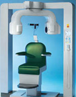
The diagnosis of intracranial hypertension and hydrocephalus is established by experts on the basis of characteristic symptoms and data of special studies, up to tomography of the brain.
It is possible to measure intracranial pressure only by administering to the liquid cavity of the skull or the vertebral channel of a special needle with a manometer connected to it. Therefore, direct measurement of intracranial pressure today is practically not applicable.
On the increase in intracranial pressure, it is safe to speak based on the following data:
- Expansion, convolutions of the Eye DNA - an indirect, but reliable sign of increasing intracranial pressure
- Expansion of liquid cerebral cavities and brain resolution on the edge of the brain ventricles, clearly visible in computer x-ray tomography (CT) or magnetic resonance imaging (MRI)
- Violation of the outflow of venous blood from the skull cavity, installed using reochephalography or transcranial dopplerography
The gold standard of instrumental examination of patients is an assessment of symptoms, data tomography of the brain and the picture of the eye don.
EchoHenecephalography (ECHO-EHO) gives indirect and not always reliable data on increasing intracranial pressure, it is less reliable than computed tomography and magnetic resonance tomography, so this method is rarely used by us.
Treatment of increased intracranial pressure
 Live with elevated intracranial pressure and unpleasant and harmful to health. The human brain under the influence of overpressure cannot normally work, moreover, slow atrophy of the white brain substance occurs, and this leads to a slow decrease in intellectual abilities, impaired nervous regulation of the internal organs (hormonal disorders, arterial hypertension, etc.). Therefore, it is necessary to take all measures to the speedy normalization of intracranial pressure.
In the treatment of increasing intracranial pressure, it is important to reduce the selection and increase the suction of the liquor. Traditionally, it is customary to appoint diuretic drugs for this purpose. However, the continuous use of diuretic drugs is not always acceptable to the patient.
Today, treatment methods are applied to normalize intracranial pressure without drugs. This is a special gymnastics to reduce intracranial pressure (applied by the patient independently), individual drinking mode and small changes in nutrition, unloading the venous bed of the head with the help of special methods of soft manual therapy.
Thus, a steady decrease in intracranial pressure is achieved without constant reception of diuretic drugs, after which unpleasant symptoms gradually decrease. The effect is usually noticeable in the first week of treatment.
In very severe cases (for example, a liquor unit after neurosurgical operations or a congenital liquor unit) applies surgical treatment. In neurosurgery, the tubing tubing (shunt) technology has been developed for removal of excess liquor.


