Magnetic resonance tomograph (MRI) is a complex system in which a powerful magnet, radio waves and computers are combined. With the help of it, the doctor receives detailed information on the location, volume and composition of the tissues of the body under study.
Content
 It is this information that can be decisive for fast, accurate and early diagnosis of diseases.
How the magnetic resonance tomograph works
X-rays are not used in MRI. As clearly out of the name, the MRI principle is to study the magnetic properties of hydrogen atoms, which are in the set in the human body. Magnetic resonant tomograph creates a strong magnetic field, which causes hydrogen atoms to fluctuate. Under the influence of radio films, atoms are slightly shifted. They are then returned to their place, and during their oscillations there is an electromagnetic signal, which can be used to reconstruct the image of the human body. Since the protons in different tissues of the human body (for example, in fat and in muscle) fluctuate with different frequencies, the image of these tissues will be different.
Contrast substances for MRI
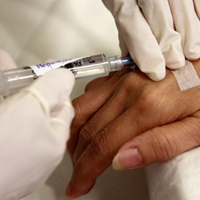
 Many modes of magnetic resonance imaging require contrast gain with special substances with paramagnetic properties. They change the electromagnetic properties of tissues and increase the contrast of the image.
Bayer Schering Pharma released the world's first contrast substance in 1988. This date has become an important event in the development of magnetic resonance imaging technologies. To date, the company has a rich experience in the development of contrasting substances for diagnostic visualization. Modern contrasting substances and special equipment for their introduction, produced by Bayer Schering Pharma, make it possible to achieve better visualization of various organs and tissues and, thus, increase the accuracy and effectiveness of medical diagnostics.
The main areas of the clinical use of MRI with a contrasting amplification of MRI is a particularly important method of diagnosing the pathology of the brain, heart, muscles and joints, as well as evaluating the state of blood vessels.
Specialized technologies of magnetic resonance imaging using contrast gain.
Magnetic resonance angiography (MRA)
Mra is used to obtain images of blood vessels (mainly arteries). This technology allows you to diagnose such pathology as an aneurysms of arteries or atherosclerosis.
Perfusion technologies MRI
Perfusion MRI is based on the introduction of the Halat of the Gadolinium and the rapid image obtaining when the bolus dose of the contrast substance passes through the blood vessels of the brain. The contrast agent causes a change in signal, the study of which in the dynamics allows you to evaluate the hemodynamics of the brain. This technology allows doctors to diagnose strokes, brain tumors and mental violations.