Benign liver tumors - these are low-power or asymptomatic formations of various nature - hemangiomas, lymphangiomas, fibromes, lipoms, Gaarterts. Read more about this in the article.
Content
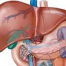
Benign liver tumors
Benign liver tumors for the majority of clinically low-alimonyptomatic benign neoplasms that occur both from epithelial tissue (hepatocellular adenoma, etc.), and from the stromal and vascular elements (hemangioma and others.).
Let us dwell on the brief description of the main ones.
Hepatocellular adenoma - A clinically small-axipty benign tumor of the type of adenoma, developing from hepatocytes, is more often graded by a capsule. With energetic growth, a tumor gap is possible with damage to vessels and bleeding.
Focal novel liver hyperplasia - Clinically small-axipty benign tumor, the central part of which is represented by a scar connective tissue, and the peripheral - nodule-transformed hepatocellular cloth. Often in the tumor there are foci of necrosis and hemorrhage. As a rule, it is not developing in a cirrotically modified liver, so sometimes referred to «Focal cirrhosis».
Promotional Regenerator Liver Hyperplasia close, and sometimes combined with focal nodular liver hyperplasia. Unlike the latter, the elements of the connective tissue are significantly lower in it. Can be considered as a hepatocellularity carcinoma. Sometimes with the growth of elements of this tumor, large bile ducts or large branches of the vein are squeezed. As a rule, it is not developing in a cirrotically modified liver.
Hemangioma liver - Clinically low-almptomatic benign tumor originating from vascular, mainly venous liver elements. Applies, apparently, to the most commonly recognized type of benign liver tumors.
All major types of benign liver tumors belong to low-voltage diseases. In many cases, their detection relates to random finds. At large sizes and the corresponding tumor localization, symptoms of biliary tract appear sometimes appear, less often - symptoms of portal hypertension.
Diagnosis of benign liver tumors
The liver is usually not significantly increased (the exception is large hemangiomas). Peripheral blood is not changed. Content of A-fetoprotein, carcinoembrium antigen, aminotransferase, GGTF, alkaline phosphatase, GDG and LDH, serum bilirubin within the normal range. Exceptions are patients who have a benign liver tumor against the background of active diffuse liver diseases.
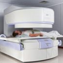
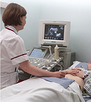
 Informative instrumental research methods. Radionuclide liver scintigraphy are performed as usual when suspected the volumetric process in the liver in two projections. With this method, you can detect a tumor with a diameter of 4-5 cm and more. In this regard, the main value of the method has in recognition of hemangiom, since the remaining three types of tumors are more often due to smaller sizes. With liver hemangiomas, 4-5 cm in size and more tumor detected in 70-80% of the surveyed. With the help of ultrasound with the hemangiom of the liver detect hyperhekogenic, well-defined education. Often, especially in the left, clearly visible vascular leg.
Differential diagnosis at this stage is primarily carried out with the parasitic liver cysts (echinococcosis). In favor of the latter, a positive reaction with a echinococcal antigen is evidenced, the katron reaction, as well as detection in the zone of tumor-shaped ordinary education.
A computer-tomographic study allows you to obtain data close to the results of the ultrasound, although it often gives additional diagnostic information regarding the first state of the surrounding tissues and organs. Colecography is most informative when recognizing hemangiom. Usually, hypervascularized areas with clear boundaries are clearly visible, allowing to detect a hemangiomic size of 2-3 cm and more than 80-85% of the surveyed.
Indirect radionuclide angiography performed using a gammakamera gives close, but less accurate compared to clerk results.
In hepatocellular adenoma, there are usually bile ductures. Therefore, when carrying out biliary radionuclide scintigraphy in the region of adenoma can be registered «Silent zones».
In the diagnosis of hepatocellular adenoma, focal nodular liver hyperplasia and nodule regenerative liver hyperplasia The results of aiming (under the control of ultrasound and computed tomography) liver biopsy play a crucial role. The complexity of the morphological assessment of the material obtained often requires the study by its morphologist and a cytologist, which specialize in the field of liver pathology.
Differential diagnosis is carried out primarily with the most frequent benign liver tumors, then with malignant tumors. In recent years, a peculiar focal dystrophy of the liver is becoming increasingly the object of differential diagnosis, especially in cases where rounded sections of intact liver are found on the background of focal fatty dystrophy. These sites have different density with fatty dystrophy and this difference is quite clearly registered with the help of ultrasound and computed tomography. These pseudo-homogeneous formations are usually not visible in radionuclide liver scintigraphy. However, this differential diagnostic sign is not very reliable. A tricky liver biopsy is playing a decisive role in identifying focal fatty dystrophy.
Treatment of difficulture liver tumors
Hepatocellular adenoma, focal nodular liver hyperplasia and nodule regenerator liver hyperplasia in medication and surgical treatment, as a rule, do not need. Exception is tumors, squeezing bile moves. In these cases, indications appear for resection of the corresponding liver segments. Methods of secondary prophylaxis. With all types of benign liver tumors, drugs such as oral contraceptives, anabolic steroids are prohibited. Unwanted reception of such drugs like phenobarbital and ziksarin. Large hemangiomes, squeezing bile ducts, remove surgically.
All patients need constant medical supervision. With the first discovered tumor, the examination is carried out after 3-6-9-12 months., Next - 1 time per year. In addition to the usual inspection of the patient with the determination of the size of the liver in Kurov, they investigate the level of bilirubin, aminotransferase, alkaline phosphatase, GGTF, GDG and LDH, A-fetoprotein and carcinoembrium antigen.