Diagnosis of lymphoma is carried out using instrumental and laboratory research methods. The main method of diagnosing the disease is biopsy. The remaining methods have additional importance when diagnosing.
Content
The diagnosis of lymphoma is carried out in several stages, the purpose of which to estimate the spread of the disease, find out in what condition the organs (liver, kidney, heart) are. The information that the doctor receives the results of inspection and examination allows it to be diagnosed, to find out the prevalence of the disease and choose the correct treatment option.
Instrumental methods of research lymphom
 If the patient has symptoms causing suspected lymphoma, you need to complete a full examination. It all starts with inspection. During inspection, the doctor carefully examines the cervical, axillary, inguinal, elbow, popliteal lymph nodes, spleen, almonds, and also explore other parts of the body to find signs that may be manifestations of lymphoma, and also learn about the status of organs about the concomitant diseases. If there is a suspicion of lymphoma, additional studies are appointed, allowing to determine the diagnosis and prevalence of the disease. These include:
- Lymph node biopsy or organ
- Abdominal ultrasound and other areas
- Chest x-ray
- CT scan
- Magnetic resonance imaging
- Radioisotope scanning
- positron emission tomography
- Blood tests - general and biochemical
- Immunophenotyping
- Survey of bone marrow
- Research of the cerebrospinal fluid
- Molecular diagnostic tests
Biopsy with lymphoma
The main test applied in the diagnosis of lymphoma is biopsy. Biopsy is a small surgical operation, during which a piece of fabric is removed (in most cases lymph node) in order to consider it under a microscope and conduct immunohistochemical, molecular and other studies. If the lymph nodes are somewhat, then remove the most changed. After a piece of fabric has been removed, it is sent to the histological laboratory for research. The information that comes after a biopsy speaks about the type of lymphoma and is key to diagnostics.
Sometimes puncture biopsy of lymph node. At the same time, after local anesthesia, the needle is introduced into a lymph node and sucking its contents. Renal diagnostics can be applied to the diagnosis of lymph in children. This is due to the fact that children are ill preferably four types of lymphoma whose cells have a very characteristic view under a microscope. The diagnosis of lymphoma in an adult is installed only and exclusively on biopsy. In most cases, children also performs the biopsy of the lymph node.
After determining the diagnosis of lymphoma, it is necessary to determine the stage of the disease, that is, finding out what other organs are involved in the pathological process.
Ultrasound procedure
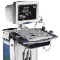
Ultrasound examination is used in the diagnosis of lymphoma very often and is assigned to almost all patients. Study based on registration of reflected ultrasound waves. It is used to find out if enlarged lymph nodes in the abdominal cavity, in the mediastinum, learn about the status of organs.
Radiography
Using X-rays, you can get a picture of the reflective state of the chest and other parts of the body. The amount of radiation that a person receives during one x-ray study is so little that you can even think about it.
Computed tomography or axial computed tomography
In computed tomography, X-rays are also used. However, pictures are made at different angles, as if around the body. Then, the results obtained are summed up into one common picture, and the computer shows a detailed shot, from each «Cut» Body. Patients with lymphomas are often prescribed computed tomography of chest, abdominal cavity and pelvis. This study is very important, it shows enlarged lymph nodes, the state of the internal organs.
Magnetic resonance tomography
Magnetic resonance tomography is similar to computed tomography. The device makes many pictures at different angles around the body, but instead of X-ray he uses a magnetic field. Magnetic resonance tomography more precisely than computed tomography. It allows you to get a more detailed picture of the internal organs, especially the nervous system. There is no more accurate way to diagnose foci in the head and especially spinal cord. It is also important in the diagnosis of bone lesions. Magnetic resonance tomography is ordered to diagnose foci of lesion in the bones, in the brain and in the spinal cord.
Radioisotope scanning with gallium
Radioactive gallium is a chemical that accumulates in the tumor. Gallium scanning is not often used in all clinics. A small number of radioactive gallium is introduced to the patient. Then the body is scanned at different angles, which would look at what places gallium accumulates. If it turns out that the tumor accumulates gallium, the scan will need to repeat after treatment. This allows you to see whether the minimum tumor remained or it disappeared at all.
Positron emission tomography
Positron-emission tomography replaced the scan with Gallium, because this technique is much more accurate. What to perform the test is intravenously introduced deoxifluoro-lucose. Then with the help of a positron camera produce the entire body scanning.
Laboratory studies in the diagnosis of lymphoma
Blood analysis
Blood test provides data on the qualitative and quantitative composition of blood. There are erythrocytes, leukocytes and platelets in the blood. Blood impairment may be the first sign of lymphoma. Biochemical blood test makes it possible to say whether the liver, kidneys, other organs are involved. Some indicators determined in the blood influence the choice of treatment and predict the forecast. For example, in patients with lymphomas, the level of lactate dehydrogenase and beta-2-microglobulin are very important, because the high level of these markers is associated with a more aggressive flow of lymphoma.
Immunophenotyping
Lymphoma cells circulating in blood or in lymph nodes are classified according to the markers on the surface or inside the cells. This method is called immunophenotyping. It is also performed on the samples of the fabrics obtained during the biopsy. Immunophenotyping results are key to lymph diagnosis.
Survey of bone marrow
Lymphoma can not only occur in the bone marrow, but also give metastasis into the bone marrow. The study of the bone marrow allows you to see, there is a defeat or not. The procedure for obtaining bone marrow is called Tpardobiopia. This is a small surgical operation. The sample of the bone marrow is taken through the thick needle of the pelvic bone, usually in the area of the belt. The procedure is performed without hospitalization and takes a few minutes.
Tpardobiopia enters the list of mandatory studies necessary to determine the stage of the disease and is carried out in most cases. Often requires two-sided trepalobiopsy, when all the same is done on the other hand.
Research of the cerebrospinal fluid
In some patients, lymphoma can spread to the nervous system. If this happens, the liquid that circulates in the spinal cord and the brain (the spinal fluid) may contain abnormal tumor cells. For diagnostics, the spinal point is performed. During the puncture, a puncture is made in the lumbar region between the proceedings of two vertebrae. A small amount of fluid is satuned in the syringe. The cells contained in the spinal fluid, as well as its biochemical composition are investigated.
Molecular diagnostic tests
Over the past decade, it became known much more about lymphoma mechanisms at molecules level, such as genes and proteins. Information obtained in numerous experiments made possible development of molecular diagnostic methods. These methods include highly sensitive polymerase chain reaction, as well as immunological methods that reveal the expression of specific proteins in tumor cells. These tests allow you to accurately determine the variant of lymphoma, follow the minimum residual disease. Information obtained using these methods allows the doctors to choose the optimal treatment to each patient. With the help of immunological and molecular methods, you can find a tumor, even in cases where the histologist does not see it under the microscope. An important advantage of molecular methods is that they are accurate and require a small amount of fabric.


