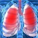
Separate types of pleurisites are characterized by a specific clinical picture. Tuberculous pleurisy can be both an independent form and a complication of the tuberculosis of another localization. Tumor pleurisy in most cases arises as a result of a metastatic lesion.
Content
Symptoms of tuberculosis pleurita
 Tuberculosis pleurisy can be an independent clinical form and as a complication of other forms of tuberculosis of any localization.
Currently, the share of tuberculous pleuritis among pleural effluents of various etiology is approximately 15-30%. Tuberculosis Pleurrites occurs in 6-8% of the newly identified patients and in 0.8-3.1% of patients with tuberculosis recurrences. Usually have young men.
The emergence of exudative pleuritis is often associated with cooling, operations, and women are still with pregnancy, childbirth and abortions. From the moment of primary fixation of the causative agent in pleural sheets to the clinical picture of the exudative pleuritis takes place from several weeks to several months. The infection reservoir can serve as intragenuous lymph nodes or residual tuberculosis changes in which the process is reactive. Extremely rarely, with an active tuberculosis of the lungs, a breakthrough in the cavity of the Caseous focus, located in the cortical part of the lung, and the development of tuberculosis empy.
Mycobacteria tuberculosis is present in excessive, which is due to the migration of them in the composition of macrophages from the inflammation zone.
Localization of pleural effusion is most often one-sided, in the lower surface departments of the chest.
Symptoms of parapneumonic pleurrites
Parapneumonic pleurisy is a complication of an intra-alert nonspecific inflammatory process (acute pneumonia, lung abscess, bronchiectase, etc.). In case of bacterial pneumonia, pleural effusion is diagnosed about 40% of patients. Most often, such pleurisy happens with pneumonia streptococcal and staphylococcal etiology. For parapnemic pleurisites, the high frequency of purulent exudates is characteristic of.
Symptoms of tumor pleurrita
Tumor pleurites in the overall structure of exudative pleurisites occupy 15-20%. The most common cause of their occurrence is lung cancer (70% of patients), lung cartridges without an established source of metastasis, mesothelioma pleura, breast cancer, gastrointestinal cancer.
The occurrence of pleural effusion depends on the nature of the tumor lesion. In metastases in the plevru, the cluster of the fluid is due to an increase in the permeability of the vascular vessels and a decrease in fluid resorption. Metastases in lymph nodes disrupt the outflow of lymph, and germination of blood vessels leads to a violation of hemocyerculation. Cancer hyphenichenemia and paracancastic pneumonia also contribute to the formation of.
With a metastatic lesion of pleura from the primary focus, located outside the lungs (for example, breast cancer), pleural effusion bilateral.
Symptoms of pancreatogenic (fermentogenic) pleurite
Pancreatogenic pleurisy is observed in 20-30% of patients with sharp pancreatitis. Pancreas enzymes come to the pleural cavity in the lymphatic vessels through the diaphragm and cause damage to the pleural sheets. Typically, pleural effusion is localized in the left pleural cavity, which is explained by the anatomical location of the talent of the pancreas in contact with the left dome of the diaphragm. In addition, it is possible to form an internal pancreatic fistula or a breakthrough of pancreatic pseudokists to the pleural cavity. In pleural exudate there is a high concentration of amylase. Exquidative pleurisy with rheumatoid arthritis develops in 4-5% of patients with rheumatoid arthritis having subcutaneous rheumatoid nodules. Usually, men are sick of such pleuris. The localization of pleural effusion is very often bilateral. When puncture biopsy parietal pleura in most cases, signs of chronic inflammation and fibrosis are found. It is possible to detect rheumatoid nodules and productive vasculitis. The flow of rheumatoid pleurite can be recurring with the formation of massive pleural overlays.
Symptoms of purulent pleuritis (Empiama Plevra)
Empiama pleura - these are all cases of pleuritis when microorganisms or pussies are found in the fluid under study. The most frequent causes of the spheres of the pleura are complicated by infectious pleurite the flow of bacterial pneumonia, subiaphragmal and pulmonary abscesses, perforation of the esophagus. About 20% of cases of emmps of the pleura are associated with spelled punings and connectible veins (with damage to the pleura). The causative agent of empya can be golden staphylococcus, a siny stick, klebseyella, intestinal wand, pneumococcus, anaerobic microorganisms.
Severe 5 groups of empya pleura:
- purulent pleurisy in the presence of a purulent inflammatory process in the body
- purulent pleurisy complicated spontaneous pneumothorax
- Piotorax, complicating therapeutic pneumothorax in patients with pulmonary tuberculosis
- Piotorax with penetrating injuries of the chest organs
- Piotorax after operations on the bodies of the chest
Pleural effusion in combination with ascites caused by non-metastatic tumors of small pelvis organs in women. The peculiarity of the tumors of the small pelvis organs is the abundant secretion of the peritoneal fluid, which, when achieving a significant volume of tumor or ascite, seeps through defects in a stretched diaphragm in the Polish cavity. Pleural effusion is located in the right pleural cavity. In the liquidation of ascites, these patients disappear and pleural effusion.


