How to diagnose an empieme of pleura? How treatment is carried out? Answers to these questions you will find in the article.
Content
Diagnostics of empya pleura
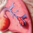
X-ray on the side of the empya is determined by intensive shading, the mediastinal displacement into a healthy side. With a popurnaceur, the upper boundary of the liquid becomes horizontal. Above the level of fluid is determined by the air cavity. Depending on the degree of lung collapse distinguish between the limited, subtotal and total pipopurnum. With limited empyrahs of the pleura to clarify the shape, sizes, the presence of bronchial fistulas is carried out by pleurography - the introduction of water-soluble contrast agents into an empieme cavity using puncture or via fistulus in the presence of a spurious fistula.
In recent years, in the diagnosis of sharp and chronic empi, the pleura is becoming increasingly distributed, an ultrasound study, especially with accused, limited empyes, when the fluid accumulations are masked by massive pleural mooring and are inaccessible to X-ray recognition. In some cases, with a deliberate purulent cavity, it is possible to perform puncture and drainage under the control of ultrasound. In difficult for diagnostics, situations primarily to identify destruction in light use computer tomography.
One of the required stages of the diagnosis of empya pleura is the diagnostic puncture of the pleural cavity. It allows you to finally establish the etiological cause of accumulation of effusion in the pleural cavity – Hemorrhagic with a lung cancer, pussy with tuberculosis, actinomycosis, nonspecific empy. Cytological, biochemical and bacteriological research of the contents of the pleural cavity is mandatory.
Treatment of empya pleura
The treatment of empya pleura includes:
- The full aspiration of the pus and the sanitation of the pleural cavity, which, with limited empiems, are achieved by regular punctures, in the aspirations of pus, rinsing the encouragement by antiseptics and the introduction of antibiotics; With open, total and subtotal empiems - drainage and washing of the empith cavity.
- Activities aimed at the early mass fraud (drainage with active aspiration, in the presence of bronchial fistula - with temporary occlusion of bronchi, or draining on Buleuu).
- Massive antibacterial therapy (cephalosporins III-IV generation, fluoroquinolones, carbapenes).
- Detoxification therapy.
- Immocorrorizing therapy.
- Lowing therapy aimed at restoring the functions of vital organs and systems.
 Local treatment of Empiama pleura largely depends on the prevalence and localization of the infectious process, the virulence of microorganisms, the characteristics of the clinical course of the disease, the presence of foreign bodies and the reports of the bronchi with pleural cavity, as well as other factors. Treatment is usually starting with suction of a pus from a pleural cavity, t. E. aspiration method. In the future, in case of insufficient effectiveness of the aspiration method, a need for surgical treatment may arise.
The aspiration of the exudate is produced either through a fairly thick needle, or through a catheter, which can be in a pleural cavity for quite a long time. It should be noted that the use of pleural punctures turns out to be effective with limited trimming empynes, preserving the tightness of the wound wound and the absence of a bronchial message with a pleural cavity. In most cases, preference is given to closed drainage of the pleural cavity without an active aspiration (siphon-drainage) or with active aspiration. This is related to the possibility of constant and more complete aspiration of purulent contents, facilities to combat local infection and the creation using active aspiration conditions for lightweight disappearances, elimination of residual cavity.
The following basic options for rehabilitation of the pleural cavity are possible: Passive drainage with fractional lavage; Permanent active aspiration with fractional lavage or continuous cavity. However, the most effective intake of treatment of acute empime is a fractional lava or continuous washing of cavity in combination with active aspiration. The point for puncture or drainage of the cavity of the pleura is determined on the basis of multi-axis x-ray. The timing of finding a drainage tube in a pleural cavity in many ways depend on the speed of the lightweight and stopping the departure of the pus, but on average, 2-3 weeks.
In the treatment of empya, the pleura is sucking if all exudate is possible. In the presence of a thick pus, fibrin films or caseous masses are used by proteolytic enzymes, contributing to the salvage of viscous pus, lysis of fibrin on pleural leaves and the potentiary action of antibiotics. Along with enzymes in the cavity of the pleura, antibiotics solutions are introduced, which are selected with the character and sensitivity of microflora isolated from exudate. In patients with acute purulent pleuritis, such treatment is usually after 2-3 weeks leads to the cessation of the accumulation of exudate in the pleural cavity, improving the overall state, the elimination of intoxication phenomena and ultimately to recovery.
If the obliteration of the lung cavity and disappearances is not achieved, chronic emmp is formed by chronic empya. In these cases, the only effective method of treatment is operational intervention - pleurectomy with lung decortication, and in the presence of destruction in light, bronchiectasis - light resection.