What is pneumophybrosis? What are his symptoms? As diagnosed and treated this ailment? Is it possible to warn pneumophybosis? You will find answers to these questions by reading this article.
Content
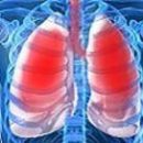
Pneumophybreas - the growth of connective tissue in the lungs due to the inflammatory or dystrophic process leading to the violation of the elasticity and the gas exchange function of the affected areas.
Prevalence can be local (limited) or diffuse.
As a rule, pneumophibrosis is the outcome of various diseases of the lungs: infectious and invasive processes (pneumonia, in T. C. Developed after mycoses, tuberculosis, syphilis, etc.); chronic obstructive diseases; Diseases caused by the impact on the body of aggressive dust and gas of industrial origin, inhalation of combat poisoning substances, hereditary diseases. Pneumophybrosis can be due to the result of exposure to light ionizing radiation, drugs with toxic effects.
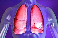
Local pneumophybosis is a plot of more dense pulmonary fabric, the volume of the affected part of the lung is reduced.
With diffuse pneumophybrosis, light dense, reduced in volume. Normal structure is lost.
Local pneumophybrosis does not significantly affect gas exchange functions and mechanical properties of the lungs. With diffuse pneumophybrosis, adequate lung ventilation decreases.
 Local pneumophybrosis clinically may not manifest.
The permanent sign of diffuse pneumophibrosis is shortness of breath, often having a tendency to progression. Often, shortness of breath is accompanied by a dry resistant cough, amplifying with forced breathing. Pains in breasts of the latter nature, weight loss, general weakness, fast fatigue. In patients with preferably damage to the basal departments of the lungs, as a rule, the so-called fingers of the hippocrates are formed (in the form of drum sticks).
Patients with pneumophybosis in far-closed stages can be detected by the so-called flaw, resembling the sound of jamming. More often it is listened to inhale, mainly over the front surface area of the chest.
The leading method of diagnosing pneumophibrosis is a x-ray study, t. To. It allows you to obtain an objective mapping of sclerotic changes of the pulmonary tissue of pneumophibrosis, to distinguish it with tumor lung lesions.
For pneumophybosis recognition, the chest air radiography is performed. Venate additions may be targeted radiography and tomography. To study the state of the lung fabric, computer tomography also acquired a special meaning.
Today, effective methods of treatment of pneumophibrosis does not exist. Local pneumophybrosis, not manifested by clinically, does not require any medicinal influences. In cases where local pneumophybrosis, which is the outcome of the inflammatory destructive process and the lungs, proceeds with periodic exacerbations of the infectious process, prescribe antimicrobial and anti-inflammatory drugs, carry out activities aimed at improving the drainage function of the bronchi affected. Bronchological research makes it possible to solve the question of the feasibility of operational intervention.
With diffuse pneumophybrosis due to external factors, it is necessary first to eliminate their impact on the patient. The treatment of respiratory failure is also carried out.
The forecast is determined by the course of the underlying disease. Formation «Cellular light» Respiratory failure sharply weights, in part of cases to an increase in pressure in the pulmonary artery system, the development of the pulmonary heart. The cause of death is often the attachment of secondary infection, the development of tuberculosis.
Pneumophybosis prevention involves the timely identification and adequate treatment of diseases leading to the development of pneumophybosis; compliance with safety regulations when working with pneumatic substances; Regular control aimed at establishing a pneumatic-toxic effect of drugs and timely conducting measures to help eliminate arising in the light pathological changes.


