How to diagnose pneumothorax? What should be emergency care for pneumothorax? Answers to these questions you will find in the article.
Content
Diagnostics
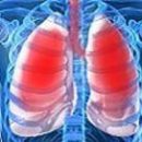
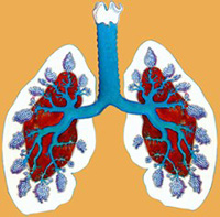
Pnemothorax
Pneumothorax recognition is easy to
The basis of acute pain in the side, increasing shortness of breath, shock symptoms,
associated with rapid air intake in the cavity of the pleura,
accompanied by a variety of pulses with pleural, lungs, small vessels
Circle, Pericarda, Hollow veins, Aorts.
Left-sided pneumothorax due to sharp pain in the area
The tops of the heart, shortness of breath, collapse accept myocardial infarction. At
right-hand pneumothorax due to the disappearance of hepatic
Stupidity, collapse may have a thought of perforation of the stomach ulcers,
12rine. Heart displacement in the other case may
accompanied by changes in the electrocardiogram similar to those that
Observed with heart attack. However, pain in spontaneous pneumothorax not
Irradiate neither in the left or right upper limbs or in the neck,
There is a unilateral absence of breathing and other signs of pneumothorax. Because of the pain in the chest, choking, collapse embolism (massive)
pulmonary artery can be mixed with suffocating pneumothorax.
Heart expansion to the right, swelling of the cervical veins, pulmonary expansion
arteries, typical steetacoustic signs of pneumothorax allow
Correct mistake. Acute respiratory failure (on the background
chronic respiratory failure) leads to an erroneous diagnosis
spontaneous pneumothorax with a sharply pronounced emphysema just like
severe attack of bronchial asthma. History, typical symptoms for
bronchial asthma and emphysema can usually establish the right
diagnosis.
X-ray study in difficult cases allows you to solve
Diagnostic task: the disappearance of the pulmonary pattern on the patient
side, pursed to the root light, in the case of fixes - a modified contour
Its, heart offset, the presence of reaching the horizontal level.
Emergency assistance for Pneumothorax
 Patient in the first hours need to have urgent
help, as it is threatened with a mortal danger. It is placed in
Bed with an elevated body position (patients themselves occupy
half-time); introduced under the skin morphine to suppress
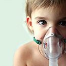
excitation, possible cough; oxygen applied against hypoxemia,
best with the help of nasal catheters connected to oxygen
Cylinder, where reducer adjusts the speed of the oxygen current. To combat S
acute cardiovascular respiratory failure
intravenous infusion of 1% novocaine solution (5-10 ml slowly for
3-5 minutes) or intravenous Introduction SOL. Papaverini 2% -2 ml. Infusion
Novocaine and Papaverin can be repeated after 4 hours. Especially
Effective wagosympo blockade on the neck (to eliminate the stream
pathological impulses with pleural and other organs) performing in
Hospital conditions.
Severe respiratory disorders when closed and
Especially valve pneumothorax are eliminated by reducing
inleptive pressure. For this purpose, they produce puncture (in
5-6 intercostal interim on the axillary line at the top edge
the underlying rib) any thick needle, be sure to
Rubber tube length in 1 m. The free end of the tube is immersed on
1-2 cm in a water vessel. The needle is removed from the cavity of the pleura only after
how the separation of air bubbles will stop through water. A place
The puncture is closed with a sterile sticker with a college. If after
pleural puncture again begins to grow shortness of breath, it is necessary
Apply constant drainage under water. In this case, the needle cannool
It is necessary to fix with leukoplast and lead for the sick
Observation.
In all cases of pneumothorax for preventing and treating infection
Plevra need to use penicillin and streptomycin in large
doses (1000000 units. - 1,500,000. Penicillina, 1 000000.
streptomycin per day).
However, with such treatment, relapses are often found, and the presence of
open fistula is associated with the risk of a pneumothorax transition in
chronic. In conditions of a well-organized hospital except
Careful x-ray examination, pleuroscopy is needed,
which makes it possible to detect the presence of pleural heavy,
Conditioning the gaping holes in Plegre. In these cases, shown
intersection of them that, of course, can be successfully implemented
Surgeon-phthisiathro.