What is traumatic synovit? How does this disease manifest? Answers to these questions you will find in the article.
Content
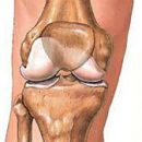
 In case of acute traumatic synotion, unlike hemarrosis, the joint increases in the amount for several hours or days. It is characterized by changing the shape of the joint, the smoothing of its contours, the increase in temperature, sickness during palpation, the appearance in the articular cavity, which is particularly well detected in the knee joint. Movement in the joint are limited, painful. There is a weakness, malaise, moderate increase in body temperature, ROE acceleration. Sinovit may complicate ankle arthrosis.
With purulent synotes, the symptoms of the disease are more dramatically expressed than in the serous. Characteristically heavy general condition of the patient (sharp weakness, chills, high body temperature, sometimes nonsense). The contours of the joint smoothed, there is a redness of the skin in the field of joint, soreness, restriction of movements, contracture. Regional lymphadenitis phenomena are often detected. In some cases, the purulent synovite process extends to the fibrous membrane of the articular capsule with the development of purulent arthritis and to the surrounding joint of the tissue. At the same time, the area of the joint is significantly increased, pastose fabrics, leather on the joints sharply hyperemic, gloms. If bones, cartridges and a binder joint joint are involved in the process, Panarthritis develops. Silver sharp synotic can recur.
Often recurrent synovites are accompanied by chronic forms of water (hydrartrosis), in which hypotrophy and its fibrosis are developing due to the constant pressure on the synovial shell, which in turn disrupts the outflow and the absorption capacity of the synovial shell. A vicious circle is formed, aggravating the synovitis and the development of degenerative-dystrophic processes in the joint.
Since the joint is a kind of organ with the specific features of exchange and livelihoods, it is necessary to dwell on the morphological and physico-chemical characteristics of the synovial medium of the joint in the norm and pathology.
The synovial fluid is normal in its composition has a significant similarity with blood plasma, as it is one of the sources of the formation of synovial fluid (synovia). At the same time, Sinovia is significantly different from blood plasma for a number of essential parameters. Thus, the content of protein in synovia is 3 times lower than in plasma, albumin ratio and globulin 3: 1, and plasma 1: 1, level α-Globulin is 3 times less than in plasma. Synovia Unlike plasma does not contain fibrinogen. Another important difference in plasma synovia is the presence of hyaluronic acid in it, which has the ability to create complex protein-polysaccharide complexes that determine the viscosity of the synovial fluid. The main source of education of the GUK and proteolytic enzymes is the coating cells of the synovial shell (synovocytes). In addition to these components, in the synovial fluid, there are numerous products of cell wear, the main substance of the synovial shell and the coating articular cartilage, entering the joint cavity in the process of life and leisis and resorption; Salts, crystals and bacteria are also present. The composition of synovia at different times and at various functional states does not remain constant. At the slightest deviation from the norm, the quantitative and qualitative state of cells, the chemical and physical properties of the synovial fluid change dramatically.
The amount of the synovia of the knee joint is low - usually 1 - 2 cm, viscosity in conventional units 5-7, pH 7.7, osmotic pressure 120-140 mm of water. Art., The number of cells of 1 cm from 13 to 200. In pathological conditions, the number of cells increases sharply and all indicators change. There is no doubt about the relationship between the total number of cells, viscosity and content in the Phagocyte synovia. Normally in the synovia cells of tissue origin prevail over blood elements (110: 100). For comparison with rheumatism (stage II), the number of cells increases sharply (1000 times or more), but also its composition varies with qualitatively: blood elements predominate over tissue cells (100: 4), and among the first neutrophils dominate (on average 68.6% by. N. Luzin, 1970). Cytological examination reveals many atypical cells uncharacteristic for normal joint synovia, as well as specific cells, characteristic of one or another pathology and the degree of development of the process (for example, phagocytes for rheumatism). Therefore, cytograms of synovia have a large diagnostic value at various pathological conditions of the joint.
Microscopically serous inflammation of the synovial shell is characterized by a pronounced vascular reaction. When synoting with the transition to a chronic form or at the initial chronic flow, the synovial shell is significantly thickened, edema, is predisposed to fibrous reincarnation. In case of recurrent synovitis, a fibrous capsule is often thickened, and a long-term synotion can lead to a joint breaking due to a sharp stretching of the capsular-ligament.