The increasing complexity of dental manipulations causes the need to master new approaches to treatment. An excellent tool that helps dentists in daily work has become a microscope. It makes it possible to more accurate diagnosis, and the use of microenvasive techniques that allow minimal injury to healthy fabrics, and the treatment of hard-to-reach areas, and control the results of treatment.
Content
The increasing complexity of dental manipulations causes the need to master new approaches to treatment. Modern technologies, materials and tools enable Dr. to consider the problem in more detail, to make treatment as accurately as possible, reduce injury and increase the aesthetic result of the work. An excellent tool that helps dentists in daily work has become a microscope. He was one of the decisive factors that changed the approach to therapeutic and surgical treatment in dentistry.
See what is hidden
There is still no unequivocal answer, who exactly came up with the prototype of a microscope - a system of convex and concave lenses in a frame capable of increasing the image, without losing clarity. Anyway, several centuries of microscopes remained the attribute of the office scientists and came into applied medicine only in the 20s of the twentieth century, when a German otolaryngologist with. Pilen offered to use a microscope during the middle ear operation. One of the first doctors, which, armed with an operating microscope, began to see what was not seen before, there were ophthalmologists and neurosurgeons. New microsurgical instruments appeared with their feed.
 In dentistry, where the main work is also conducted with small and very small objects, the lenses began to work in the 50th. Microscopes came to the US dentists in the USA, in the 80s, when medical practice has already accumulated sufficient experience in microsurgery. And it produced a real revolution. «The appearance of such optical devices can be compared with the situation when a physician who acts on the touch suddenly appeared vision, "approves Kristina Stryapin, Therapist «Professor's dental clinic», - Tactile sensations added the opportunity to see the area of work. Due to this: the amount of information that the doctor receives in diagnosis has increased, the quality of treatment has increased, the possibilities for the treatment of diseases in the early stages have increased».
«Professor's dental clinic» One of the first in Russia introduced a microscope in his practice. Grigory Drobot, To.M.N., Chief Doctor, Surgeon «Professors» It says that she felt the hard share of pioneers in itself: «One microscope is not enough, you need skilled hands to manage them. And at first it was possible to learn this only abroad. Even now, I can not confidently say that a high-quality system of working with a microscope has been formed in Russia. That is why our doctors are still involved in international seminars and master classes, and now, often, and as teachers», - Notes O.
Upon closer look
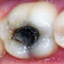
Of course, the main value of the microscope is the possibility of a multiple increase in a small object or its elements. Dentistologists of all specialties «Professor's dental clinic» There are incredible advantages that makes it macroevitis - it is the possibility of more accurate diagnosis, and microenvasive techniques that allow minimal injury to healthy fabrics, and treatment of hard-to-reach areas, and control of the results of treatment. For the patient, all this means only one thing - he is becoming more and more chances to keep a brilliant smile to deep old age.
Zries in the root
 The coup of the microscope produced in therapy and endodontics, the most important part of practical dentistry, which is engaged in the treatment of pulp diseases. Elena Haustova, To.M.N., Therapist, head of the therapeutic department «Professor's dental clinic», 6 years ago, one of the first in Russia has mastered the technique of working with a microscope in dental practice. According to her, he turned out to be an indispensable tool for a doctor. «Now it is even not clear how it was possible to work without a microscope using the light of the ordinary dental installation, and search channels, based on its own intuition», - Remembers Elena.
The microscope, in addition to the knowledge and shape of the channels available to the doctors, gives a specialist qualitatively new information. For a doctor, dentin under the microscope works as a card with which it will see the secrets of the tooth anatomy and will detect the hidden mouths of the channels. This is especially important when treating the tooth, poorly treated earlier. In the process of treatment, again with the help of a microscope, the doctor will make sure that all the remnants of the old sealing material have been carefully removed, the lively and healthy tiss fabric is most as possible, and the new materials tightly filled the cavities without leaving air pockets for bacteria. «Simply put, the microscope gave me and my colleagues in the clinic the opportunity to not only represent, but also to see all the anatomical features of a tooth, but, it means to treat it, and not some imaginary object», - Hears Elena Haustova.
Jewelry work
No less decisive role microscope played in dental surgery. The term of the new century has become «Microinvasia» - minimal surgical interference in the modified, patients of the tooth, allowing to save the tooth until it is possible in principle. Grigory Drobot says that dental operations are comparable by subtleties with neurosurgical: «In the operations on the brain, during all sorts of transplants, visualization plays a huge role: the two ends of the vessel should be connected absolutely accurately with the smallest number of seams so that the postoperative period is successful and did not arise the thrombus». In dental surgery, everything is also happening - with a microscope, a doctor can more accurately compare the edges of the wound and avoid complications and scars on the mucous membrane.
With the help of macro-milling surgeons were able to carry out operations with maximum accuracy, use microsurgical instruments, reduce the trauma of interventions, reduce the postoperative period and increase the aesthetic result of the work. «With dermal operations, such as resection of the root tips, some periodontal operations, the microscope is simply necessary - continues Grigory shot. - On the tooth and inside it there are cracks and fractures that are visible only with increasing. Previously, such operations were intuitive, with some number of unknown, and their success was usually 60 - 70%. Microscope allowed us to bring the percentage of successful operations almost up to one hundred».
The accuracy with which allows the operation of the microscope, played its role in this direction of dentistry as «Pink aesthetics». Her predecessor - «White aesthetics» - This state of the crown of the tooth. Doctors have long learned to make a crown of natural and transparent. «But alone she is nothing without beautiful, healthy gums. It is comparable to a expensive stone that requires a decent frame», - Explains the term Grigory Drobot. Now in dentistry they received the distribution of a correction operation or an increase in the volume of gums to make it harmoniously looking. «This is a simple operation, but it must be done very carefully and carefully, accuracy and minimum number of seams are important in it, because, firstly, we are talking about aesthetics, and secondly - in the operation zone there must be sufficient blood supply to the operation. Microscope allows you to achieve», - Stresses Surgeon.
A look into the future
 The design of dental microscopes has changed the work of doctors and their assistants physically. The changing angle of the eyepieces, the possibility of rotating the design allows the doctor to perform painstaking and long work in a convenient position, sitting or standing, without excess voltage in the muscles. Focusing and towing fields occur in a fraction of a second, and the specialist is not distracted from the patient to set the eyepiece. The system of live broadcast of the manipulation that the doctor produces helps video assistants to navigate in the operating field, to properly file the desired tool at the right time.
Christina Stryakina marks another huge advantage of a microscope - this is the ability to display an image on the screen, write a video, take a photo. «It incredibly helps us in dealing with patients», - She notes. Patients interested «Professor's dental clinic» not only know what happens to them, but can also become eyewitness. Video and photos are archived and persist in the electronic history of the disease, which even after a few years will be able to any doctor quickly and clearly describe the patient's condition and show the process dynamics, make an idea of the predisposition to one or another diseases and facilitate the preparation of the treatment prediction.
In Russia, microscopes are just starting their way in dentistry. But it is known exactly exactly the future. An example of this is the radical changes that made microsurgery into ophthalmology, and obvious changes in dental practice abroad. After all, according to one of their first doctors, applied a microscope in Dentistry Harry. Carr: «You can not treat what you do not see».