The number of pregnant women with heart disease has recently increased. To this group of diseases include heart defects. Thanks to the improvement of methods for diagnosing and treating heart disease, many «Hearts», previously doomed to infertility, got the opportunity to endure and give birth to a child.
Content
The concept of heart disease
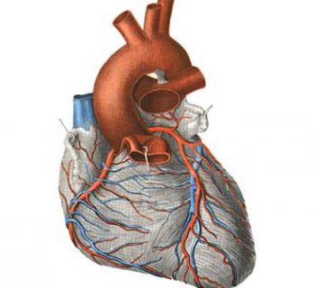
The normal operation of the heart largely depends on the functioning of its valve apparatus. The defects of the heart are, first of all, a violation of the work of cardiac valves (folds, opening and closing holes between heart chambers, as well as between heart and large vessels; the correct operation of the valves provides blood circulation).
Heart Pulk can manifest itself in the form of valve deficiency (valve flaps do not close to the end, and the reverse blood flow occurs) or stenosis (narrowing) of the hole when the blood flow is created. Often stenosis and insufficiency develop on one valve (the so-called combination vice). In addition, there are cases when the defects affect two or more valves - it is customary to be called a combined defect of the heart.
When heart disease, blood circulation is broken. This is especially dangerous during pregnancy, when the load on the cardiovascular system increases.
Heart defects are congenital and acquired. Almost all acquired heart defects are developing against rheumatism, they may occur during pregnancy (the aggravation of rheumatism in pregnant women is most often observed in the first three and the last two months of pregnancy). Most often on the background of rheumatism, combined defects of the mitral (located between the left atrial and left ventricle) valve are developing, less often - the aortic valves. The most frequent and serious vice - mitral stenosis (that is, the narrowing of the hole of the two-hearted heart valve).
Diagnostics of heart defects
Currently, there is a wide arsenal of diagnostic methods and treatment of rheumatism. Women suffering from rheumatism, it is especially important to plan a pregnancy. A favorable forecast of the course of pregnancy is possible if it has come against the background of an inactive rheumatic process.
Modern medicine has fairly effective techniques that allow calculating the degree of risk associated with pregnancy and childbirth in women with heart defects. With their help, doctors help a woman to determine the optimal time to conceive or solve the fate of unplanned pregnancy.
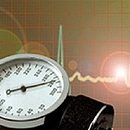
The most important methods for estimating the condition of the cardiovascular system at heart disease is the ultrasound of the heart - echocardiography. It is harmless and helps objectively assess the condition of cavities, valves and heart openings. An auxiliary role in the diagnosis of heart defects is played by electrocardiography (ECG - graphic registration of electrical activity of the heart), phonocardiography (FKG - graphical registration of heart sound phenomena) and Doppler (ultrasound, allowing to appreciate blood flow).
Currently, the opportunity to give birth to many women who have undergone heart surgery. The recovery period after such an operation takes, as a rule, 1 year. Therefore, one after a year it is possible to plan a pregnancy - of course, in the absence of contraindications (the unfavorable result of the operation, the development of diseases complicating postoperative rehabilitation and reduce the effect of the operation). It is too easy to remind that the question of the possibility of pregnancy and the admissibility of childbirth should be solved individually before pregnancy depending on the general state of the woman, the nature of the disease, the severity of the operation and T.NS. After a comprehensive examination of the patient, the doctor can give a certain conclusion.
However, when stabilizing the state of a woman after surgical (or therapeutic) treatment, pregnancy against the background of the growing load on the heart increases the risk of recurrence of the main disease (a previously compensated vice can become decompensated) - this is another argument in favor of the need for consultation with a doctor and medical observation before and in pregnancy time, even if the woman itself it seems that she is healthy and full forces.
There are severe heart defects with significant circulatory disorders (stenosis of the mouth of the pulmonary artery, Tetrad Fallo, coarctation of aortic and others .), in the presence of which can develop so dramatic disorders of the cardiovascular system, which in 40-70% they lead to the death of a pregnant woman, so with these vices, pregnancy is contraindicated. Such vices can be inherited, and the probability of transmitting the disease to the child is determined in each case.
In general, the forecast for the future mother and the child is the worse, the more pronounced circulatory disorder and the activity of the rheumatic process. With severe heart failure and high degree of activity of the rheumatic process, pregnancy is contraindicated. However, the question of pregnancy pregnancy is solved by a patient and a doctor in each case individually.
Features of pregnancy at heart defects
 During pregnancy, the load on the cardiovascular system increases significantly. By the end of the second trimester of pregnancy, the blood circulation rate increases by almost 80%. The volume of circulating blood also increases (by 30-50% to the eighth month of pregnancy). This is understandable - after all, the blood flow of the fetal is joined to the parent circulatory system. With such an additional load in a third of pregnant women with a healthy heart, impaired cardiac rhythm (arrhythmias) and the work of the heart valves, what to talk about women with heart defects.
If necessary, drug treatment with heart defects is carried out throughout the entire pregnancy. The purpose of treatment is the normalization of blood circulation and the creation of normal conditions for the development of the fetus. Cardiac glycosides, anticeriotic products, diuretic, vasodilators, antiagregants and anticoagulants are used (means that prevent blood coagulation and thrombus formation). The question of the appointment of drugs and their doses is solved individually, in accordance with the term of pregnancy and the degree of severity of circulatory disorders. In the ineffectiveness of therapy resort to surgical treatment, preferably on the 18-26 week of pregnancy.
Throughout the pregnancy, cardiotokography (ultrasound of the fetal heart) is performed periodically. With the help of dopplerography, the uterine-placental and fetal (fruit) blood flow is investigated to eliminate hypoxia (oxygen starvation) of the fetus. Naturally, constant control over the state of the mother's heart is carried out.
Often, even with initially compensated vice during pregnancy, complications are possible, so each pregnant, suffering from heart disease, should at least three times during pregnancy pass to examine in cardiology.
The first time - on the period of up to 12 weeks of pregnancy, when after careful cardiological and, if necessary, rheumatologic, surveys, the issue of pregnancy is resolved. The second time - in the period from 28 to 32 weeks, when the load on the heart of the woman is especially large and it is very important to carry out preventive treatment. After all, a large load on the heart can lead to development at this time:
- Chronic heart failure characterized by fatigue, edema, shortness of breath, increased liver
- Heart rhythm violations (arrhythmias)
These complications may occur not only during pregnancy, but also in childbirth, and in the early postpartum period. For a child, such violations of maternal blood circulation are fraught with a lack of oxygen (hypoxy). If you do not accept timely measures, a delay of intrauterine development may be observed, insufficient body weight (hypotrophy) of the fetus.
The third hospitalization is carried out 2 weeks before delivery. At this time, a re-cardiological examination is carried out and a manual plan is produced, preparations for them.