Decoding ultrasound results. The essence of the method, which signs of the disease can identify a diagnostic.
Content
Ultrasound Research Method (ultrasound)
Ultrasound Research (Ultrasound) — This is the safest method of visualization of pathology located in any organs of the human body.
The essence of the method
The study is based on the irradiation of human body with ultrasound waves and registering them. Any fabric, except bone, misses ultrasound. On the border of tissues with different density there is a partial reflection of the rays that are fixed by the sensor. The information received is processed by a computer and is issued on the image as an image.
Decoding results
Decoding the result of the ultrasound is largely subjective and depends on:
- equipment on which research is carried out;
- Professionalism of the doctor;
- the correctness of the preparation for the ultrasound (for example, before the study of the bladder should be drunk waters so that it is filled).
In the study, the doctor may reveal:
- Signs of inflammation — thickening of the walls of hollow organs, an increase in the echogenicity of parenchymal organs;
- Availability of neoplasms — cyst, tumors;
- presence of stones in some organs — liver, kidneys, bile tracks;
- Changes in large joints — the state of the ligaments, cartilage, the volume of the joint liquid;
- Changes from large vessels.
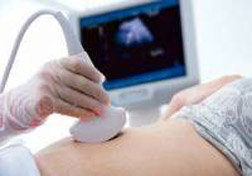
In pregnant women, this method is a mandatory screening element — Periodic control. It is used for:
- definitions of pregnancy;
- Floor definitions of the fetus;
- detecting various pathologies of placenta and uterus;
- early detection of disorders of the development of the fetus.
At the ultrasound of patients, a doctor can direct any specialty, ranging from the district therapist and ending with the neurosurgeon.