Find out to which doctor to appeal to decipher the results of vascular ultrasound; What diseases may evidence vascular ultrasound and types of ultrasound research.
Content
Decoding the results of tests of vascular ultrasound
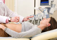
Ultrasound examination (ultrasound) vessels allows determining the condition of the structures of the circulatory system, which supply the brain with blood, neck, upper and lower limbs, sex, pelvic organs and kidneys.
Types of ultrasound vessels
- Doppler ultrasound determines the rate of reflection of ultrasonic waves from erythrocytes and gives a two-dimensional image showing the presence or absence of vessel narrowings.
- Duplex ultrasound — Combination of Doppler ultrasound with traditional — gives a color image, shows the violation of the normal course of blood flow, its speed and direction.
- Triplex ultrasound differs from duplex multicolored blood flow velocity.
Duplex ultrasound examination determines:
- violations of cerebral circulation (occlusive diseases of the carotid arteries);
- Raino diseases, top breast aperture syndrome and veins thrombosis — We are talking about the problems of the vessels of the upper limbs;
- Lonic atherosclerosis, arterial aneurysms and deep veins thrombosis, varicose veins — diseases of the vessels of the lower extremities;
- Aortic aneurysms (abdominal and thoracic departments);
- narrowness of aortic and iliac vessels.
Ultrasound safe and helps in the diagnosis of pulsating formations and observing patients with coronary heart disease.
Preparation for duplex ultrasound scanning is necessary only in the case of aortic (abdominal vessels): It is necessary to put an intestine to purify the intestines.
Obtain an appointment for testing vessels by neurologist, cardiologist, fluball, surgeon and therapist.