Acute leukemia is a severe blood illness at which an uncontrolled reproduction of immature blood cells occurs, which, unlike mature, is not able to perform their functions. How can this disease can be diagnosed?
Content
Is it possible to early detection of leukemia
Currently, there are no special methods to diagnose acute leukemia at an early stage. The best recommendation is an urgent appeal to the doctor when any inexplicable symptoms appear. People who are in high-risk groups should be under regular and thorough observation.
How acute leukemia is diagnosed
Leukemia can be accompanied by many signs and symptoms, some of which are nonspecific. Note that the symptoms below are most often happening with other diseases, and not when cancer.
 Common symptoms for leukemia may include increased fatigue, weakness, weight loss, increased temperature (fever) and loss of appetite.
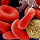
Most of the symptoms of acute leukemia are caused by a decrease in the number of erythrocytes as a result of the replacement of the normal bone marrow, producing blood cells, leukemic cells. As a result of this process, the patient decreases the number of normally functioning erythrocytes, leukocytes and thrombocytes.
Anemia (Malokrovia) is the result of a decrease in the number of erythrocytes. Anemia leads to shortness of breath, fatigue and skin pallor.
Reducing the number of leukocytes increases the risk of developing infectious diseases. Although patients with leukemia, the number of leukocytes can be very high, these cells are not normal and do not protect the body from infection.
Low platelet count can cause bruises, bleeding from nose and gums.
The spread of leukemia beyond the bone marrow to other organs or the central nervous system can cause various symptoms, such as headache, weakness, cramps, vomiting, violation of gait and vision.
Some patients may complain about pain in the bones and joints due to their leaning leukemic cells.
Leukemia can lead to an increase in the size of the liver and spleen. With damage to lymph nodes, they can be increased.
In patients with acute myeloid leukemia, the lesions of the gum leads to their swelling, soreness and bleeding. Lesion of the skin is manifested by the presence of small multicolored spots resembling rash.
When the T-cell type of acute lymphoblastic leukemia is often amazed for a fork gland. Large vein (top hollow vein), carrying blood from the head and upper extremities to the heart, runs next to the fork iron. Increased forks of the fork gland can squeeze the trachea, causing cough, shortness of breath and even suffocation.
When squeezing the upper floor of the veins is possible swelling of the face and upper extremities (the top hollow syndrome of the vein). It can break the blood supply to the brain and be life-threatening. Patients with such syndrome should start treatment immediately.
Methods of diagnosis and classification of leukemia
The presence of some of the above symptom does not mean that the patient has leukemia. Therefore, additional studies are conducted to clarify the diagnosis, and in confirmation of leukemia - its type.
 Research of blood. Changing the number of different types of blood cells and their appearance under the microscope can give reason to assume leukemia. In the majority of patients with acute leukemia (acute lymphoblastic leukemia or acute myeloid leukemia), for example, there are too many leukocytes, few erythrocytes and platelets. In addition, many leukocytes are blast cells (the type of unripe cells, in the normal non-blood circulating). These cells do not perform their function.
Survey of bone marrow. Using a thin needle, a small amount of bone marrow is closed for research. This method is used to confirm the diagnosis of leukemia and evaluating the effectiveness of treatment.
Biopsy of the lymphatic assembly. With this procedure, the lymphatic node is removed and then investigated.
Spinal brain puncture. During this procedure, a thin needle is introduced in the lower back area in the spinal channel to obtain a small amount of spinal fluid, which is studied to detect leukemic cells.
Laboratory research. Various special methods are used to diagnose and refine the type of leukemia: cytochemistry, flow cytometry, immunocytochemistry, cytogenetics and molecular genetic studies. Experts study bone marrow, lymphatic tissue, blood, spinal fluid under a microscope. They estimate the size and shape of cells, as well as other cell characteristics to determine the type of leukemia, the degree of cell maturity.
Other research methods
- X-ray pictures are performed to identify tumor formations in the chest cavity, bone and joint lesions.
- Computed tomography (CT) is a special method of x-ray research, allowing to examine the body at different angles. The method is used to detect the lesion of the chest and abdominal cavities.
- Magnetic resonance imaging (MRI) uses strong magnets and radio waves to obtain a detailed image of the body. The method is especially justified to assess the state of the head and spinal cord.
- Ultrasound (ultrasound) allows you to distinguish tumor education and cysts, as well as the state of the kidneys, liver and spleen, lymph nodes.
- Scanning of lymphatic and bone systems: with this method, the radioactive substance is introduced intravenously and accumulated in lymph nodes or bones. Allows differentiate between leukemic and inflammatory processes in lymph nodes and bones.