What to do in erosion of the cervix in pregnancy? Why appears erosion during pregnancy? Read in this article.
Content
Colposcopic picture
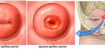
 Colposcopic pattern of the mucous membrane of the vaginal part of the cervix in a healthy woman is different in different days of the menstrual cycle.
In the first phase – This is a smooth surface of light pink color, not significantly changing after processing by mucous painshing solutions
In the second phase, when the surface layer of a multi-layer flat epithelium occurs, the mucous membrane still remains smooth, pink, but additionally vessels begin to visualize on it. This is understandable, the layer of a multi-layered flat epithelium thinned. These vessels, after using coloring solutions, give a characteristic picture called papillary relief.
Changes on the mucous membrane part of the cervix, as mentioned above, are associated with fluctuations in the level of sex hormones in the body of a woman (the predominance of estrogen in the first phase of the menstrual cycle and progesterone in the second).
And now imagine how the concentration of hormones should change during pregnancy, considering the placenta – This is a kind of temporary endocrine organ in the body of a woman. Naturally mucous membrane of the vaginal part of the cervix will not remain unrequited for such hesitations.
Musocolism
In the first trimester of pregnancy, the mucousa acquires a blue color. The hyperplasia of a multilayer flat epithelium is occurring, the differentiation of cells is often disturbed, a large number of cells in mitosis appears. The mucous membrane of the cervical canal thickens, the cells of the cylindrical epithelium increase and all this is accompanied by the strengthening of the mucus separation. Hypertrophies and backup cells.
In the II and III trimesters, due to the increase in the cervix, the boundary of the junctions of the epithelium can shift towards the vaginal surface of the cervix, T.E. Physiological ectopia is formed.
As an option of the physiological norm in a small number of women on the mucous membrane, areas of the growth of decidual tissue can appear (literal translation - dropping fabric), called decophyte (growing outward) of cancer with a naked eye when inspection).
Localization of dezidosis may be different: in the field of external parasis, vagina arches, in the cervical canal and T.D.
Colposcopically decidulose is determined very easily, and considering the literal translation of the dezidosis, as a disappearing fabric, it disappears spontaneously after delivery.
However, remember that even in the physiological course of pregnancy and colposcopy, and cytologically can detect some atiypius cells, which makes it difficult to formulate the correct diagnosis.
Atipia
No need to be scared «Atipia» and take it immediately for cancer. Atipia – it is only nonypical cells, whose cells have any changes. And how I have already noted above: «In the first trimester of pregnancy……and T.D.», T.E. The body is preparing for childbirth, cells change to perform their functions well during pregnancy and childbirth and atypius (atypical cells) in this case – it's not cancer, it's a normal condition.
But since changes in the human body can not always be predicted with 100% accuracy, and in pregnancy, the diagnosis is generally difficult, so it is necessary to conduct dynamic colposcopic observation with the mandatory use of cytological, and if necessary and histological research methods.
Only treatment does not need any, dezidosis is not treated, it is a physiological norm. What the norm is to treat, and even more so caress – freeze? Like ectopia without signs of atypics.
So, future moms, do not worry if the gynecologist, with the usual inspection, will fix you «country-a-ahnyh» Shake changes. Perform colposcopy, and everything will fall into place.


