What is an intracranial hypertension syndrome? How it manifests and how to treat the sick? Read in this article.
Content
Intracranial hypertension syndrome
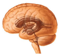
Intracranial hypertension syndrome — One of the most common brain damage clinics is due to the excess accumulation of the spinal fluid (liquor) in the ventricles of the brain and under its shells arising from the obstacle to the outflow, redundant education and disorders of the inverse absorption of liquor. In order to better understand the essence of this process, let's dwell on the anatomy (structure) of the brain and the mechanisms of fluid outflow from it. The human brain has several cavities called ventricles (large 4), they are interconnected and filled with a special liquid (liquor), which is produced by special structures — Vascular plexuses from blood flowing in the arteries. Then the lycvore is absorbed into venous vessels, replacing new. The brain, given its high need for oxygen, needs enhanced blood supply, therefore, blood flows into it in four major arteries, and it reaches the veins. For the proper functioning of the brain, a good movement of the ceremony lycvore is required and between the shells of the brain, good absorption of it in the venous network and the outflow of blood from the brain on the veins.
In violation in any link of liquorodynamics, the outflow of excess liquid is hampered, it accumulates in the ventricles of the brain, expanding them between the shells. Veins are overwhelmed with blood, and the child of the first year of life increases not only the size of the brain ventricles, but also the size of the head. Great spring increases in size, empty, pulsates, siggital seams diverges, but it is precisely it helps the child for a long time to compensate for excessive accumulation of fluid.
Hydrocephalus — This "dropsy" brain, excess liquid. Hypertension — This increases pressure resulting from a fluid pressure on the brain substance. Both components are closely interrelated, and have a common name — "Hypertensional-hydrocepal syndrome".
Causes of occurrence
It should be noted that this syndrome may occur as due to organic brain damage ("Mechanical" blockage of liquid outflow tumor or hematoma) and inorganic lesions associated with a decrease in the vascular tone, in particular venous, which leads to the difficulty of the outflow of excess liquid and overflow of the ventricles of the brain.
Manifestations of intracranial hypertension syndrome

Clinically hypertensive hydrocephalic syndrome is manifested by the following signs: the children of the first year of life are sluggish, weak, suck well and quickly get tired; Frequent vomit "fountain"; shrill crying, more similar to moan; tension, sprouting springs; the discrepancy of the cranial seams; an increase in muscle tone in the limbs, especially in the legs; convulsions; Atrophy of optic nerves. In children older than the year (with closed spring), signs of intracranial hypertension can develop very quickly, they are manifested by strong, parotid headaches, more often in the morning, with vomiting that does not bring relief. The behavior of children is changing, at first they are restless, any external stimulus is annoyed (bright light, loud sound and so on), then children become sluggish, larger. Sometimes there is a fixed position of the head, an afflicted facial expression. On the eye day there are stagnant phenomena, a decrease in visual acuity.
It should be noted that in children of any age there may be so-called transient (transient), fluctuations in likvorn pressure. Headache, nausea, dizziness and other symptoms can be a manifestation of both a set of functional impairment of brain activity and various tumors (both benign and malignant), abscesses, hematomas, infectious and other diseases. Depending on the causes of the occurrence of hypertensive-hydrocephalic syndrome, there will be different and treatment — From drug, aimed at improving fluid outflow, to a surgical, eliminating cause of occlusion (blockage) of a liquor outflow.
In order to establish the true cause of hypertensive-hydrocephalic syndrome, it is necessary to conduct a comprehensive clinical examination of the child.
Children of the first year of life, the monthly increase of the head of the head (in children of the first half of the first half, on average, 2 cm per month, in children of the second half of the year — 1 cm per month). For comparison, I will give a few numbers: the normal circle of the head at the ended healthy child is 34-36 cm at birth (usually the boys are 1-2 cm more than girls), in 3 months — 38-42 cm, in 6 months —42-46 cm, at 12 months — 46-48 cm, in a year and a half — 46-50 cm, in 2 years— 48-52 cm.
Here it is necessary to separately dwell on the group of children who have somewhat circumference slightly exceeds the average age norms, and in the absence of any signs of lesion of the brain, you can not worry. Children who have suffered in the first year rickets, as well as children whose parents have such a constitution — so-called hereditary macro — It is necessary to conduct differential diagnosis with hypertensive hydrocephalic syndrome, and in the absence of ventricular expansion, treatment is not required.
Additional research
 To clarify the causes of the disease, it is necessary to carry out the following hardware examination:
- EchoHehephalography (ECHEG) — The method of diagnosing intracranial lesions with ultrasound does not have contraindications, provides high accuracy, can be applied in children with almost birth;
- Reoencefalogram (REG) — Explore the venous outflow of brain vessels is carried out in children from birth;
- Radiography skull — more informative with a long time of the current disease, more often used in children older than one year;
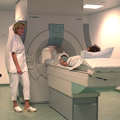
- Computed tomography (CT) — Allows you to accurately determine the plot of occlusion of liquorottock, the size of the ventricles and so on;
- Electroencephalography (EEG) — Determination of brain activity processes using electrical pulses.
There are inspections by such specialists as an ophthalmologist, a neuropathologist, a psychiatrist (long-term hypertensive-hydrocephalic syndrome can lead to atrophy of the cerebral cortex and, subsequently, to a delay in mental, mental development), neurosurgeon.
Treatment
Depending on the reasons that caused the disease may be drug (dehydration therapy with diaklab, in combination with loopholes, massage and physiotherapy) and surgical (removal of education that interferes with fluid outflows or, if it is impossible to carry out such an operation, shouting of brain ventricles — Shunt is inserted — Special tube), with the help of which the lycvore recesses from the ventricles of the brain directly to the lower part of the spinal canal.
Hypertensional-hydrocephalic syndrome is rarely isolated, and often combined with oppression syndrome or comatose state, consider their manifestations.
Syndrome inhibition
The syndrome of oppression is manifested by lethargy, hypodynamines, a decrease in spontaneous activity, total muscle hypotension, oppression of newborns, reduced sucking and swallowing reflexes. This syndrome characterizes the course of an acute period of perinatal encephalopathy and by the end of the first month of life usually disappears. But it may be a forerunner of the brain edema and the development of comatose syndrome.
Comatous syndrome
Comatous syndrome is a manifestation of the extremely heavy state of the newborn (on a scale of apgar such children have 1-4 points). In the clinical picture, lethargy, adamope, a decrease in muscle tone to atony, innate reflexes are not detected, the pupils are narrowed, the reaction to the light is minor or absent at all, "Floating"The movements of the eyeballs, there is no reaction to pain stimuli. Breathing arrhythmical, with frequent apnea (stop), bradycardia (heart rate gentle), heart tones Deaf, pulse arrhythmic, blood pressure low, no sucking and swallowing reflexes.
This state of the newborn requires urgent therapy and is extremely uncertain in terms of development and health of the child.


