What is hemangioma liver? What is hepatoenoma? How to diagnose and treat these tumors? Answers to these questions you will find in the article.
Content
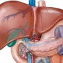
Hemangioma liver
 Hemangioma can be solid and multiple (10%),Turning with hemangiomas of other organs, more often have a cavernous structure. By origin, they are related to congenital diseases. Describes seMeyan cases of illness, women suffer 5-7 times more often than men. With a microscopic examination, a rich network of vascular lacuna with thin connective tanksMi partitions. In rare cases, hemangioma can reach considerable sizes, occupying the right or left lobe of the liver.
Clinical picture and diagnostics. Gemangiomas of small sizes (meIt is 5 - 8 cm in diameter) Usually BessemPtomns. The disease usually exhibitat the fourth-fifth decade of life when reached the tumorSize. For all kindnessTyphic Typical Stupid NoahPains in the right hypochondrium, less often - symptoms of internal organs (stomach, duodenum). In an objective study of patients with large hemangiomas and other tumors, hepatomegaly (increase in liver) is detected, significantly less often can be placed and a tumor dropped during palpation, sometimes above the zone of proHemangioma levion of systolic noise. When gigantic hemangiomas, heart failure may develop due to massive blood discharge through arteriovenous anastomoses of this tumor.
Among the instrumental ways to study the most informativeIt is a computed tomography at which a rich liquid is detectedbone multi-chamber tumor; celiacography; Ultrasonic research; laparoscopy.
It is possible to develop such complications as the gap of hemangioma with profuse (often fatal) intra-abdominal bleeding; thrombosis of vessels feeding the tumor, followed by necrosis (less often with abscedation) of the tumor; Rarely - Malignization (Acquisition by cells of the properties of a malignant tumor).
Treatment. With small (less than 5 cm) hemangiomas surgical treatmentNot shown, it is necessary to dynamic observation. Large heman-genomas that threaten the development of complications, it is advisable to delete - withChange the resection of the liver, the volume of which is determined by the geeman-genoma, or the so-called near-tumor resection (essentially enuclayaHemangioma).
Hepatoenoma
Hepatoenoma (developing from hepatocytes) and cholangogeatom (oncevisiting the epithelium of bile ducts) are rarely found, they are usually asymptomatic. Clinical manifestations of the disease occur only when the tumor of large sizes, when bleeding, or when twisting the tumor located on the leg, in connection with the node necrosis. Basic SPODiagnostic EY - Ultrasound Research and Computer Tomography. Hepatomas are sometimes soligrated, so it is appropriate toTop (enucleation, wedge-shaped liver resection). Cholangogeatoms are not prone to malignancy; For small sizes of the tumor and the absence of clinical manifestations, their removal is not necessary.
Other benign liver tumors of mesenchymal and mixedorigin is extremely rarely found.