Complications for hemangiome liver. Methods of diagnosis and treatment of hemangioma.
Content
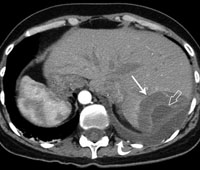
Hemangioma liver — This is benign vascular
tumor, which is detected by about 7% of people. Usually does not have a tendency
to malignancy (reincarnation in malignant state), women meet
more often than men 3-5 times. With small sizes, the tumor does not represent
Danger for life, while growing up to sizes Over 3 cm Requires treatment.
Hemangioma liver is a benign
Vascular neoplasm Diembormbrioplastic or Blastomatous.
Typically, the size of the tumor does not exceed 3 cm, in rare cases can reach 5-6
cm.
Causes of occurrence and types
tumors
Exact causes are not established. It is believed that the disease
can develop as a result of a congenital defect and after receiving drugs on
The basis of estrogen.
Views
Two types of tumors distinguish:
-
Capillary hemangiomas — Consist of small
cavities in size 2-3 cm in diameter. Often each of them has a separate vein. -
Cavernous hemangiomas — Consist of small
cavities that are combined into large. Have uneven contours and inhomogeneous
structure, their size can reach 20 cm.
Symptoms
With small neoplasm sizes, the disease proceeds
Asymptomatic. Symptoms begin to manifest when the neoplasm reaches enough
big sizes and begins to influence other organs. Usually
There are noving pain in the right hypochondrium, sometimes — feeling
comprehensive internal organs, nausea and vomiting are possible. Due to squeezing
The growing tumor of the branches of the vein inside the organ and bile mainstone
The tributaries can manifest the symptoms of portal hypertension and obtuctive
Jaundles.
Complications
In the case of a large tumor with a non-time
The development of various complications. Among them: Heart
Insufficiency, thrombosis of the supply vessels and tumor breaking with profuse
intra-painted bleeding capable of leading severe bleeding and
fatal outcome. Tumor break is rare, but dangerous complication,
Therefore, in the event of acute abdominal pain, urgent
hospitalization of the patient.
Diagnostics

In suspected a disease, a general blood test is carried out,
and the patient is sent for further examination. The most common method of diagnosing hemangioma in state hospitals — Ultrasonic
Studying liver. Modern methods usually apply in private clinics,
allowing you to put more accurate diagnoses — Computer and magnetic resonance
tomography. In suspected the development of hemangioma of the right lobe of the liver Patient
directed to angiography, allowing to find out the condition of blood
Vessels. During hemangioma is prohibited biopsy, since the procedure causes
bleeding.
Treatment
If the tumor of small sizes and its presence is no
danger does not represent, then treatment is not required. But in the case of growing it
may cause serious consequences.
Operational intervention
With the accompaniment of the growth of complex symptoms, it is shown
Surgical Operation on Complete Tumor. Surgical intervention
required if the tumor reached more than 5 cm in diameter, is located
superficially squeezes internal organs and rushes a threat to infection or
rebirth in malignant. Operation is contraindicated in lesion
The main veins of the liver and with a passing discovery in the patient of the liver cirrhosis. Not
A hemangioma is also removed simultaneously on both fractions of the liver.
Non-surgical methods
Often practiced non-surgical treatment with
using a laser, microwave radiation, radiolochy therapy, electrocoagulation and
Cryodestruction. During radiotherapy, atypical cells provoking
Development of the disease, using laser technologies sclerosized
Affected vessels. The basis of cryodestruction is the use of
Low-temperature liquid nitrogen, and electrocoagulation — High frequency
Electric current. In drug treatment, hormonal is carried out
Therapy, the duration and dosage of which is determined individually.