Teratoma does not spare neither adults or children. Since this tumor grows from the embryo cells, then the children are subject to it at least than adults. Read more about teratoms in childhood - read in the article.
Content
 Theratomas are found mainly in children of the first years of life, less often - in adolescence and make up about 6% among children's tumors.
Newborn and infants with tumor diseases of teratomas are found in 22-25% of cases.
Places of accommodation by teratom in children are distinguished by a variety: a sacker-cork region - 38%, ovaries - 31%, retroperitoneal space - 12%, eggs - 6%, mediastum - 4%, other - 9%.
The origin of the tertol today to the end and not studied. Some of them are vices of the development of the nucleus tissue, others - the result of the wrong development of one of the germons of twins, solded together.
In newborns, teratomas are usually built of immature fabrics and are difficult distress from malignant tumors. In children aged 4 months to 5 years, teratomas are malignant in 50-60% of cases.
With the external arrangement of the teratoma in the sacrochikovsky area, it is easily detected at birth, as it is located on the buttock or by the middle sacring-smoking line.
In the external internal location of the teratoma, except for the serving tumor, there is another part located between the sacrum and the rectum.
With the growth of the inner part of the teratomas, the symptoms of the disorder of pelvic organs in the form of urination disorders, constipation or incontinence. Such a teratom is often a packer soldier, grows the dust and can be accompanied by a leather decay, an attachment of infection and bleeding.
Tematoma to the touch can be smooth or buggy (with malignant reincarnation), leather over it - unchanged or necrotic (dead) at large tumor sizes. Tumor consistency with a benign teratoma is usually softer than with malignant. Leather temperature over malignant teratoma is usually increased, and the vascular pattern is well designated. Unlike benign teratom, malignant varieties of tumors rarely reach large sizes.
With benign teratoms, the overall condition of the child suffers little, if the tumor does not squeeze the surrounding organs and fabrics. Local symptoms are always pronounced in children with malignant tumors. In addition, there is a pallor of the skin, weight loss, a lag in physical development, an increase in temperature, an increase in ESO (erythrocyte sedimentation rate). Children are often restless, which is associated with pains, and which are indicated by older.
Theratoma of ovaries is a long time flows. The first sign of the disease is usually an increase in the size of the abdomen. Sometimes children complain about pain at the bottom of the belly. Twisted the legs of the beaters of the ovary can cause, except for pain, vomiting and the tension of the muscles of the front abdominal wall (the so-called syndrome «acute belly»).
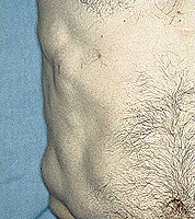
During the retroperic teratoms, symptoms appear caused by squeezing of the surrounding organs and tissues: periodic pains, extension of blood vessels on the front abdominal wall, the presence of a tumor when tamping belly.
In patients with theratoms of external localization (neck, egg, etc.) There are usually local symptoms.
In the radiography of the chest can detect the metastatic lesion of the lung fabric.
Radiography of the spine allows you to be distinguished by a tremble vertebrae, leading to the spinal hernia.
With a radiographic study of the teratoma, various inclusions can be detected in it.
Ultrasound examination (ultrasound), computed tomography (CT), magnetic resonance tomography (MRI) make it possible to establish a tumor connection with surrounding tissues.
In suspected of involvement in the tumor process of large vessels, angiography is used (contrasting of vessels), and if the bones are suspected, radioisotope scanning of the bone system is performed.
If possible, the tumor puncture is made to determine the degree of maturity (malignancy) of the process.
Determining the level of alpha-fetoprotein (AFP) makes it possible to clarify the malignant nature of teratoma, as well as monitor the effectiveness of treatment.
 Treatment with teratom in children is based on the degree of maturity (malignancy) of the tumor. With mature (benign) teratoms, the optimal type of treatment is a radical operation with a tumor removal along with its capsule in extremely early time.
In children with immature (malignant) teratomas, a combined (combined) treatment is applied. The implementation of radical operations with immature teratoms is difficult due to the increase in tumor and its local and remote distribution (metastasis).
The radiation method of treatment with immature terats is usually applied as an addition to a non-radical operation. It should be noted a minor sensitivity of malignant tertot to irradiation.
In the case of incomplete removal of immature teratoma and the presence of metastases, chemotherapy of vinnicistine, dactinicine, adriamycin, cyclophosphamph, bleomycin, semeside, methotrexate or platinum preparations can be applied. In some cases, there is a reduction in the size of the tumor and metastases.
Survival in children with immature terators is much worse compared to patients with mature tumors, and largely depends on the radicality of operational intervention (completeness of removal).
After the completion of the entire treatment program, children should be under constant observation of oncologists. This is due to the fact that with incomplete (nera-s) removal of the tumor is possible recurrence (refund) of the disease. This is especially true of patients with malignant teratoms, when, after incomplete removal or inadequate treatment, there may be relapse of the tumor in its primary location or remote metastases.


