Did you think about how the skin is arranged, liver or spleen? And how is our stop? Only when they are beginning to disturb pain, discomfort, millions of questions appear. What hurts? Why hurts? All these bones and tendons exist? Together with you we study the anatomy of the foot
Content
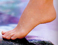
Foot – This is an organ with a very difficult anatomy. There is a fairly large load on it. She participates in the act of walking and therefore its condition is reflected on our gait.
Anatomically in the foot allocate the following areas:
-
Front surface
-
Rear surface
-
Side surfaces: Medial and lateral.
In addition, the sole and the back surface of the foot stands out.
Sole of the foot – The most important part in terms of the gait and state of foot joints. The soles are allocated vaults: longitudinal and transverse. In this case, the internal longitudinal arch of the foot has supporting structures:
- Long and short plantar bundles,
- Fastened fascia
- fifty.
Violation of the supporting function of these components leads to such pathology as flatfoot. These structures are under the influence of some factors weaken and no longer withstand the load that the stop carries.
Bones Foot
 Bone «Frame» Foot consists of 26 bones. At the same time, several departments stand out in the bone skeleton of the foot: it is repulse, plus and phalange of the fingers.
Tarsus – This is a foot department, which is located between the tie bones and ankle joint. It includes the following bones: tan, heel, lands, lateral, intermediate and medial wedge-shaped bones and cubic bones. Tweets – Five bones that belong to tubular bones.
Falanga – short tubular bones of which are formed. The thumb form forms two phalanxes, the remaining fingers are formed by three phalanxes. Among the joints of the foot is the main one, the main place is occupied by an ankle joint. It is formed by the tibia shin and football feet. The remaining joints are small (tall-standing, interfalane and joints are repluted).
Tweets carry the main load with the heel bone.
Heel bone There are some feature – on its plantar surface can sometimes form a thickened thigh – «heel spur». In terms of pathology it is important to note the first advantage of the inflave joint. Most often in this area marked arthrosis (degenerative diseases of the joint).
In addition, the area of the head of the first tie bone plays a special role in the emergence of the Valgus deformation of the thumb (Hallux Valgus). This is manifested by the fact that bone outflow is formed on the side surface of the head of this bone. He squeezes soft fabrics in this place, and also causes the deformation of the joint, which is manifested by pain and disruption.
Bundles and tendons
The bones of the feet are connected with each other with ligaments that strengthen the joints. The most important bundle in the foot is a deltoid bunch. It is located on the inner (medial) foot surface and connects the tibia, tanning and heel bones. A very important role is a binding device playing in maintaining foot arches.
Bundles of foot Traumatization is often subject to: breaking and dumping. Note that a fairly frequent term «Stretching» can not be attributed to bindings, as the ligaments are a rather strong structure. Therefore, when injured, they usually observe their gap or, more often – Partial supervision.
Tendon – This is what the muscles are attached to the bones. According to their structure, they resemble ligaments. Both ligaments and tendons are formed from collagen fibers, which, as it were, woven in the form of a rope. It provides them with strength and definite elasticity.
The most famous tendon on the foot is Achillovo tendon. It is attached to the heel bone and is a continuation of the icy muscle. It participates in the flexion of the foot.
Foot bones, connecting with each other, form a joint. Each joint is surrounded by a joint capsule, which is strengthened by bundles. From the inside the body cavity is covered with a synovial sheath. When injury to large joints – in this case ankle – Blood can accumulate in his cavity. Hemarthrosis is formed.
Muscles of the foot
 Muscles answer all the listed foot structures. Muscles on the foot are distinguished long and short. Long muscles originate from the bone of the leg, from its top. Short muscles begin with lower shin departments.
All foot muscles are divided into flexors, extensors, as well as inter-care and black-shaped muscles. In its location, they are divided into the muscles of the stalls of the feet and the muscles of the sole.
Muscles of the foot are driven by nerves. Among the nerves that are responsible for the movement of the foot – Tibra and Deep Malobers. Skin sensitivity on the foot corresponds to the surface small-terror nerve, as well as the skin branches of the above two nerves.
The blood supply to the foot is carried out with the help of two arteries: the front and rear tibial. Front tibial artery forms a so-called arterial arc on the rear of the foot. The rear tibial artery goes on the sole of the foot and is divided into two branches.
Having given in the fabric all nutrients and oxygen and taking slags and carbon dioxide, blood from the foot reaches two veins: large and low subcutaneous. At the same time, a large subcutaneous vein goes along the inner surface of the foot, and a small – by outer.