Overview of irrigoscopy
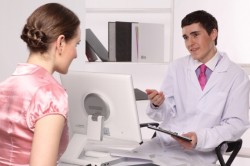
This examination is carried out using X-ray rays, while X-ray-repeat substance is used. The method allows you to consider the area that is presumably susceptible to illness. During the procedure, the doctor has the opportunity to make several pictures. But at the very beginning of the procedure, the patient in the anal hole is injected with barium sulfate (these contrasts) or combine the barray with air (this is already double contrast). The length of the large intestine in person from 1.2 to 1.5 m, it consists of several departments that perform their function. The method will show the state of the descending, rising departments, the rectum, separate sections of the small intestine and Appendix (blind process). The ability to stretch the intestinal wall will be determined. If there is disorders of the ileocecal valve (it is located between the ileum and the colon), it will also be detected. It is possible to obtain data on the mucous membrane and the activities of different intestinal segments. Irrigoscopy allows you to clarify the diagnosis, and the patient does not spend a lot of cash and time. The motor activity of the intestinal apparatus will also be studied, and this is especially important if the patient has a chalk problem.
When irrigoscopy is shown?
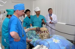
The intestinal irrigoscopy can be carried out only in those medical centers and clinics where there is special equipment. A device that allows for the procedure is called Bobrov apparatus. At first, the medical staff connects a contrast agent with ordinary water, while observing certain proportions. After the solution heated to 35 degrees. It will be introduced to the patient rectally (in the rear pass) using the Bobrov apparatus. The heated composition is poured into a special capacity of this device, it closes with a lid to which two tubes are attached. At the end of one of them there is a rubber pear required to injected air into a vessel. At the end of another tube there is a replaceable tip (it happens different sizes). This device allows you to enter the barium in the intestine. To make sure that there are no roam masses in the intestines, first the patient will be made x-ray. And only then a solution containing an X-ray-repeat substance will be introduced. Volume can be from 1.5 to 2 liters. Snapshots will be made several (they are called visible and aimed), all of them are conducted under different angles, so the patient will be asked to lie on the belly, on the back, on the side. At the final stage, a snapshot of the entire intestine will be fully filled with barium. It will analyze the form of this body and find out the distance of the lumen. The final snapshot will be completed after the patient is emptying the intestines. If the intestinal walls should be considered in more detail (with suspected the presence of ulcers, polyps, cancer), then the patient make double contrast. To do this, use a device that allows me to be supplied to feed the air flow into the intestine to fix everything in the intestine.
The duration of such a survey in some patients is 10 minutes, others - 30-40 minutes. The examination patients are transported calmly, noting only the fact that when they are poured into the intestines of the barium composition, they feel a call for defecation, small spasms and gravity in the abdomen.
After examination, doctors will certainly advise the patient in the following days to take more fluids to quickly remove the barium composition from the intestine. Coming home, immediately athe. Elderly patients are preferably in bed on the first day. In case of difficulties with the emptying of the intestine, you can resort to an easy laxative preparation.
results

With a healthy intestine, the doctor will see that he will dissolve evenly, physiological protrusion is present and clearly designate. As soon as the intestine is emptied, it acquires a normal state, and the mucosa has a peristically.
With inflammation (colitis, diverticulosis), the doctor will determine the locations of their localization and the extensity of the plots.
With tumors (sarcoma, adenocarcinoma), these formations are similar to the snapshot on the grizzle from the apple, they narrow the intestinal interlock, while the intestinal barium is filled with an abnormal manner. However, 100% confirm the presence of irrigoscopy tumor still can not. To confirm the diagnosis, it will take biopsy, endoscopic examination.
Contraindications to irrigoscopy

The doctor will not be able to give an appointment to this x-ray examination, if the patient has the following diseases and symptoms:
- acute diarrhea, with the presence of blood impurities;
- If there is a risk of intestinal spreading;
- Megacolon (vice, in which the large intestine is increased);
- Swift ulcerative colitis;
- tachycardia.
The procedure is not carried out by pregnant women. There are doctors who believe that this examination is better not to perform offen women.
There are relative contraindications:
- Acute form of intestinal supply of blood.
- The probability of presence in the intestine of cystic pneumatosis.
- Acute course of processes in the colon.
Do not be scared, if in the first three days after such a survey in your felling you will see whitish impurities - this is increasing the radiopatrum. In rare cases, symptoms may appear to alert:
- stomach ache;
- bleeding from the rear pass;
- vomiting, slide;
- dizziness, weakness;
- temperature increase.
Noticing such signs of unfavorable with health, do not hesitate to immediately contact the doctor.

