Any disease is most successful in the initial stages. In relation to such a not gone as keratoconus this statement is more than right. That is why it is very important in time to recognize this twig. How diagnosed keratoconus is diagnosed, as well as its causes and forecast, the author of this article knows.
Content
Keratokonus: It is important to recognize on time
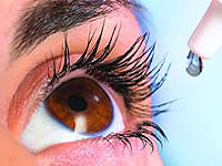
 Keratokonus - eye disease, in which the cornea is thinning and takes a conical form.
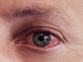
Keratoconus can lead to serious impairment of vision. Most often, patients complain about the lights, bone, image wrapping.
Symptoms of keratoconus and diagnosis
Ophthalmologist or Okulist usually proceeds to diagnose without the use of special tools. He talks with the patient, paying attention to the main complaints and subjective symptoms of violation of vision, possible injuries or diseases capable of damaging the eyes, and the family history of eye diseases. Then uses a visual check table.
 Another feature can give a procedure at which the doctor directs a ray of light on the patient's rainbow shell, and monitors the reflection, shifting the beam. Keratoconus and some other diseases create the so-called «Effect of scissors», When the two reflected stripes of light move to each other and back, as if the teeth of scissors.
In case of suspected keratoconus, the doctor conducts an inspection of the cornea with a slit lamp. If the disease has already developed enough, such an inspection allows you to immediately make a diagnosis without resorting to more specific tests.
More accurate diagnostics ensures the topography of the cornea, at which a special device projected on the cornea is analyzed by a computer to calculate the topology of its surface.
The topographic map reflects all the irregularities and scars of the cornea, and the characteristic increase in curvature is clearly seen with keratoconus. This is especially important for the early diagnosis of the cornea, when other signs have not yet appeared. Comparing several topographic snapshots, you can estimate the nature and rate of corneal deformation.
Causes of keratoconus
Distortion of view arise for two reasons - due to the deformation of the cornea and due to the border of its surface. The curve surface breaks the image into parts, leading to the symptoms of monocular polyelation, deteriorating in the dark when the pupil expands and opens to the review a larger area of the cornea.
Some researchers suggest that, for some reasons, free radicals and other oxydant substances accumulate in the cornea. Obviously, regardless of the cause caused by the cornea damage leads to its thinning and mechanical weakening.
Genetic predisposition to the occurrence of keratoconus is proved in family studies and studies of single-square twins. Most geneticists converge on the fact that the disease is inherited. People with Daun's disease more often than others sick keratoconus, but the reasons for this correlation are unknown. Keratoconus is accompanied by diseases such as asthma, allergies, eczema, and often a person is striking at once all of these ailments. A number of studies suggests that excessive eye friction by hands accelerates the course of the disease, and patients should avoid physical impact on the eyes.
Forecast with keratoconus
Keratoconus in most patients occurs during the start of puberty in the form of weak astigmatism and is properly diagnosed only after a while. The disease rarely occurs in adults, after puberty or in children. Early keratoconus is associated with a greater probability of severe course. The visual acuity changes in the better, then for the worst side for months after the start of the disease, forcing the frequent change of glasses. Keratoconus is diverse - in some patients the course of the disease stops for many years, others have a rapid drop in view, the third phases of stability are replaced by a jump-shaking acceleration of illness.
In severe cases, the foul in the cornea can lead to the local rupture of its inner layer. The patient feels pain and sudden bolding of the field of view, and a milky white spot appears on the cornea. This phenomenon is called «Water catch cornea». Despite soreness and inconvenience, the transparency of the cornea usually returns six to eight weeks later. You can speed up the recovery process using osmotic saline solutions.
In particularly severe cases, a partial rupture of the cornea occurs, and a small swelling of the amount with a bead occurs on its surface. There is a threat to increasing the break and loss of the eye. In this case, the donor corneal is carried out by an emergency transplant.