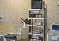
The main methods of diagnostics of endometrial hyperplasia are separate diagnostic scraping of endometrials followed by histological examination. Histological research is the most reliable way to diagnose the disease.
Content
Histological methods of study of endometrial hyperplasia
The main diagnostic method is the separate diagnostic embodiment of the endometrium followed by histological examination of the material obtained.
The scraping of the mucous membrane is carried out either on the eve of the alleged menstruation, or at the very beginning of the appearance of bloody discharges. In the diagnosis of endometrial hyperplasia, endometrial scraping should be extremely carefully, with the removal of the entire mucous membrane. Such a need is caused by the fact that the foci of adenomatosis and the polyps are often located in the region of the bottom of the uterus and the uterine pipe corners.
The uterine mucous membrane removed as a result of the scraping is directed to histological examination, that is, the study of organism tissues. During histological examination, a morphologist, given the data on the age of the patient, its complaints, the main symptoms, the nature of the menstrual cycle, the duration of the disease, the clinical diagnosis, evaluates histological data.
Histological research is currently the most reliable method for diagnosing endometrial hyperplasia and determining its form (iron-cystic hyperplasia, atypical hyperplasia is represented by adenomatosis diffuse, focal, polyps may be fervor, adenomatous, fibrous).
For more accurate diagnostics, endometrial scraping can be conducted under the control of hysteroscopy, that is, using a special magnifying device called a hysteroscope.
The use of hysteroscopy in the diagnosis of endometrial hyperplasia helps to study the endometrium state in more detail and monitor the results of the therapy.
After the scraping made, its quality is also estimated using a hysteroscopic study, it allows you to remove the residues of hyperplasted endometrial or polyps, identify the accompanying intrauterine pathology.
Liquid hysteroscopy, which is used most often, allows you to perform a number of intrauterine operations, use electric and laser surgery.
In order to monitor the treatment of endometrial hyperplasia, as well as with a dispensary examination of a woman, this method is also used to diagnose as an aspiration (that is, suction using special instruments) of the uterine cavity content with subsequent cytological examination.
Aspiration is carried out in compliance with the rules of aseptic, in the second half of the menstrual cycle. This survey cannot be used to determine the nature of the endometrial hyperplasia, its ability is limited to the determination of the presence or absence of pathology.
Ultrasound Methods
 An important role in the diagnosis of endometrial hyperplasia is currently played by an ultrasound study (ultrasound) with a vaginal sensor, which allows you to determine the endometrium hyperplasia by the nature of echo signals.
In particular, the presence of endometrial hyperplasia may indicate the endometrium thickness, which is easily determined when ultrasound. As for the polyps of endometrial, then with ultrasound, they are visible on the echoogram in the form of rounded or elongated oval formations with a clear contour and a thin echonegative rim against the background of an extended uterine cavity.
Finally, such a method for diagnosing endometrial hyperplasia is more and more important is the radioisotope examination of the uterus.
With this diagnostic study, it is possible to determine not only the presence of hyperplastic processes in endometrial, but also the degree of their activity.
For radiometric research, indicator doses of radioactive phosphorus are usually used.
Careful diagnostic examination, as a rule, includes a whole range of diagnostic methods. With the modern development of medicine, endometrial hyperplasia is diagnosed quite easily, and therefore the likelihood that the patient will be fully and quickly cured from the unpleasant consequences of this pathology.


