In the modern world, the gall-eyed disease is quite wide, and the symptoms of the disease are known to most women. What is required for the correct diagnosis and is it worth referring to the doctor when the disease is first manifest?
Content
The first symptoms of cholecystitis - reason to appeal to the doctor
 If for a long time you are borrowing in the mouth, nausea, pain and severity in the right hypochondrium, arising after meals (especially after consuming oily food, fried meat, smoked, salted, pickled products, wine or vodka), it's time to go to the reception to Doctor-therapist. His task is to figure out what is happening with you, because with diseases of the gallbladder, symptoms are the most different, nonspecific. This means that such pains or nausea can cause ulcers, pancreatitis, liver disease. And determine the diagnosis of gallway disease «On the eye» hard even an experienced doctor.
Therefore, additional analyzes are often required to confirm the diagnosis of gallstone disease. If necessary, the therapist will send you to a gastroenterologist who is engaged in the treatment of uncomplicated diseases of the gallbladder and bile ducts. And only in the absence of the effect of therapeutic treatment or development of dangerous complications of complications for the case, surgeons are taken.
Main complaints in chronic cholecystitis
On the primary reception in your interests as you can read your complaints. For example, if the pain is worried, the doctor will be interested in:
- where and when hurts
- What is the character of pain (it is constant or grapple)
- What makes relief (perhaps it is a hot tub, antispasmodics)
- What aggravates pain (it can be shaking, oily or fried food)
- Whether vomit happens
- Have you noticed darkening of urine and discolored feces
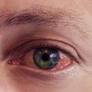
- Is there no shifts of eye or skin proteins
Characterize pain after she has already passed, not always easy, so it's worth writing something right away as soon as you feel able to do it.
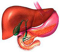
Then the doctor must inspect you. How do you know the gallbladder is right under the rib arc. The doctor especially carefully trucks this zone, as well as other stomach areas. It is important to inspect the eye proteins to notice the early manifestations of jaundice. However, with the disease of the gallbladder outside the exacerbation, as a rule, a person looks quite healthy. That is why in most cases additional analyzes and surveys are appointed to confirm the diagnosis.
Laboratory research methods
Blood tests. The doctor may assign blood test on transaminase. This analysis refers to functional hepatic samples (that is, it shows how the liver works). If the stone has blocked bile moves, transaminases can be elevated. Clinical blood test is also made. If the number of leukocytes is increased, the gallbladder is possible.
 Abdominal ultrasound. Most of you are familiar with this procedure, which is held on an empty stomach. A transparent gel is applied on the stomach, which facilitates the penetration of ultrasonic rays through the skin. Using a special sensor, ultrasonic rays are sent to internal organs. The sensor moves across the abdomen, and the doctor sees the image of the organs on the screen. If there are stones in the bustling bubble, they are well noticeable. Ultrasound gives an accurate result in the diagnosis of gall-eyed disease in 95% of cases, which is quite good. Using the ultrasound, you can also measure the dimensions of the gallbladder and the diameter of the ducts; see if there is an inflammation of the wall of the gallbladder. Other abdominal organs are also being investigated: liver, pancreas, spleen, kidneys.
Cholecystography of gallbladder. Before the ultrasound began to use, most often resorted to cholecyistography. If this examination is prescribed, you need to swallow several tablets of the contrast agent that will go into the gallbladder. After some time, X-rays do. If there are stones in the bustling bubble, they will look like «Defects filling», that is, the plots not filled with a contrast. However, if the gallbladder is amazed, the contrast agent may not go into it.
Gallbladder scintigraphy is a very accurate and expensive view of the survey, which is carried out in specialized clinics. The doctor may appoint it if he believes that your complaints are associated with a gallbladder disease, and ultrasound and cholecystography did not give a diagnostic result. Scintigraphy - radioisotope examination, but the dose of irradiation is extremely small, and it is absolutely harmless. A small amount of radioisotope drug is introduced into Vienna. It is captured by the liver and accumulates in a bustling bubble. The doctor by scanning determines the degree of this accumulation. If the accumulation does not occur, possibly bubbled bubbling. If the mucous membrane of the gallbladder is damaged, it can not be visible at all. According to the results of scintigraphy, the question of the removal of the gallbladder is often solved.
Biliary gallbladder. This is an even more special study, which is carried out only in specialized gastroenterological clinics. It is intended for measuring pressure at the end of the biliary tract, the sphincter is apparently. Sometimes this sphincter is spasked, and the bile cannot act in the duodenum. When this happens, the patient has pain, liver samples change.
Retrograde cholangiopancratography (RHPG) is assigned if the patient complains of pain in the right hypochondrium, and other studies, such as ultrasound and computed tomography, do not allow to establish an accurate diagnosis. The study is carried out on an empty stomach in the X-ray department. The nurse will handle the rear wall of the pharyx with an anesthetic aerosol so that its sensitivity decreases. A special soothing remedy will be introduced into the vein so that the patient relates to, but was in full consciousness. The doctor then cautiously introduces a catheter in the esophagus and then through the stomach - in the duodenum. He will bring it to the fare of the nipple, where the duodenum opens a common bull duct. Next, the catheter is carefully introduced into the overall biliary duct through the panels of the nipple, and a contrast agent is supplied under low pressure through it. Then X-ray shots are made and the state of bile and pancreatic ducts is estimated.
Duodenal sensing. This study used to be used often, and now it is not enough informative. A probe is introduced into the duodenum, then the gallbladder is stimulated with medicines and take a few bile samples in different moments of bile intake in the duodenum.
As you can see, doctors have a whole arsenal of diagnostic research, but in most cases, blood tests, ultrasound and sometimes additionally cholecystography make it possible to diagnose the gallway disease. When it is necessary to insist on hospitalization, and when you can be treated in the clinic? It is impossible to give recommendations for all occasions, so let's consider the most typical situations.