What surveys are necessary to confirm the sclerosis? Read in this article.
Content
Diagnostics of multiple sclerosis
The diagnosis of multiple sclerosis is primarily based on the availability of typical symptoms, which are features of this disease, as well as on the results of the visualization of the brain.
Analysis that would allow immediately determining the presence or absence of multiple sclerosis until there is. None of the existing tests that help doctors when making a diagnosis, in itself is not 100% convincing.
The neuropathologist will establish a final diagnosis by eliminating other possible reasons:
Asking questions related to the history of the disease, the doctor will determine whether you have noted any neurological disorders that could be so insignificant that you may not have taken them seriously.
Conducting a thorough neurological examination, the doctor will pay attention to the presence of signs and symptoms of neurological changes.
Collecting additional data, the doctor is looking for reinforcing facts to clarify the diagnosis. These data are obtained from various sources, such as MRI shots (Magnetic resonance imaging), laboratory tests (oligoclonal bands), tests for assessing the electrical activity of the brain (caused by potentials) and, finally, studies of the spinal fluid (lumbar puncture).
Neurological examination with multiple sclerosis
Through a neurological examination, it is checked as far as the nervous system of man works. Ordinary neurological surveys are usually carried out by a neurologist. Neuropathologist will check the presence of any changes in visual and hearing perception, as well as changes in sensitivity and violation of speech.
In addition, it is likely to also check the reflexes of elbows, wrists, knees, ankles and soles of stop to find signs of another possible violation
Monitoring and walking (t.E. For such parameters like foot movements, posture, crawls with hands with standing or walking, the presence of weakness or spasticity of the muscles) may reveal an ataxia or loss of sensitivity reflecting damage to nerves in the dorsal or brain.
The doctor can also check the presence of changes affecting ears (hearing), face (loss of sensitivity), throat (notch), as well as.
The optic nerve, leading from the brain to the eyes, is often amazed at multiple sclerosis. Spectator nerve surveys include visual testing, visual caused potentials, as well as a thorough examination of the eye itself.
Vision is checked using ophthalmological tables, and the optic nerve is examined using an ophthalmoscope. All these checks are simple and painless.
Revealed pathologies, such as eye twitching (nystagm), can reflect the activity of multiple sclerosis or to detect the existence of other violations arising in the field of the inner ear.
Along with a neurological examination, additional diagnostic procedures are carried out in the standard order, such as obtaining pictures magnetic resonance tomography. Despite the fact that this is the only study in which you can see the foci of multiple sclerosis cannot be considered final. Scanner can show not all foci, especially in the early stages of multiple sclerosis, and, moreover, some other diseases detected by the method magnetic resonance tomography, Can cause identical changes in the nervous system.
Magnetic resonance imaging
 Magnetic resonance tomography (MRI) allows you to clearly define the size, quantity and distribution of foci or plaques in the brain and in some cases in the spinal cord.
Magnetic resonance imaging is a very useful tool due to its ability to demonstrate changes in the activity of multiple sclerosis over time.
The detection of foci at the early stage of the inflammatory process can be improved by introducing a contrast agent into a vein in the area of the axillary depression or forearm.
Snapshots magnetic resonance tomography allow you to visualize the foci-related sclerosis. Along with reinforcing facts obtained when studying the history of the disease and neurological examination, the foci, which are detected when magnetic resonance tomography , are an extremely significant indicator of multiple sclerosis.
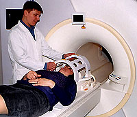
Magnetic resonance imaging is a highly sensitive technique used to determine the foci in the brain. The process of obtaining a picture MAnti-resonance tomographyand can be quite tedious for a person who makes a tomogram, because it will have to spend some time. Obtaining a conventional picture may take 10-20 minutes. All this time, the patient will have to lie absolutely motionless on the table that moves inside a wide pipe. This procedure can be quite noisy and cause a sense of some shy.
In many medical centers, the patient will be able to take advantage of earplugs or listen to music during the procedure.
In addition, the patient will supply the emergency call button to communicate with the radiologist while in the tomograph pipe.
X-ray radiation magnetic resonance tomography scanning is not used, so this survey can be repeated as often as it will take. With additional questions should be addressed to your doctor.
Caused by cerebral potential
With multiple sclerosis, signals on various nerves slow down as a result of damage to the nerve fibers of myelin shells, insulating and protecting these fibers. On such sites «Golden» nerves transmission of impulses is significantly delayed.
When measuring induced potentials, it is necessary to accurately fix the time for which some irritant reaches the brain. Delays are detected by comparing the results of the survey with the amount of time, which usually takes the transfer of pulses in people who do not suffer from multiple sclerosis.
When measuring the electrical activity of the brain, neuropathologists can detect foci that do not cause the appearance of clinical symptoms. Non-invasive techniques, such as caused by the potentials reflecting the brain electrical reaction to a number of stimuli, are still value, especially with doubts about the diagnosis.
Cropped potentials can help neuropathologists to detect disorders of nervous conductivity and so-called «silent» The foci in the central nervous system is already when no neurological deficit is not yet observed.
Caused potentials are used not only for the diagnosis of multiple sclerosis, they are also important indicators of the course of this disease.
Summary caused potentials
Visual caused potentials are most often analyzed as part of the diagnosis of multiple sclerosis. The time is measured, the required visual nerve for the transmission of visual information into that section of the brain, which primarily is responsible for processing such information.
The patient's head is placed electrodes that fix electrical activity (brain waves) in the brain. To measure pulses passing by a visual nerve between the eyes and the brain, the patient will be asked to concentrate its attention on the screen resembling a chessboard, with a small square in the middle.
Damage to the optic nerve can determine the pathological indicators. Consequently, the identification of such deviation in a person with clinically normal vision can help confirm the diagnosis of multiple sclerosis,.
Laboratory research
Spinal Puncture / Lumbar Punch
 Diagnose scarm sclerosis is not easy, therefore, additional data obtained in laboratory studies may be useful. One of the tests is the fence of the cerebrospinal fluid (SMG) from the liquid-containing spinal portion. Liquid extracted using the needle when the patient is in the lying position. For this procedure, it may be necessary to stay in the hospital for several hours.
Spinal puncture applies to confirm or eliminate the diagnosis of multiple sclerosis. With the spinal fluid (SMG), a number of analyzes can be carried out, which can identify the availability of the activity of multiple sclerosis.
Most people with mounted sclerosis (90%) have a positive result of the analysis of SMI for the presence of multiple sclerosis.
In general, nor the results of manimal resonance tomography, neither caused by the potentials nor the spinal points themselves do not diagnose multiple sclerosis, t.E. A reliable diagnosis of multiple sclerosis cannot be based only on these studies. These studies are primarily a subsidiary to establish a final diagnosis, helping to confirm or eliminate the suspect diagnosis, so each of these analyzes must be carefully interpreted by an experienced specialist.
Diagnosis at earlier stages of the disease
Various techniques have been developed that doctors are guided in their diagnosis. To establish a reliable diagnosis of multiple sclerosis, certain diagnostic criteria must be performed.
Today, modern methods magnetic resonance tomography And these new criteria allow doctors better differentiate multiple sclerosis from other diseases in which similar symptoms may be present.
Thanks to these advanced diagnostic techniques, attending physicians today have a much higher chance to start an effective treatment of multiple sclerosis in the early stages of the disease. Early therapy will reduce or derete the risk of irreversible damage to nerve fibers in the future.


