What kind of specialization doctor can put or clarify the diagnosis «Hemangioma»? What means of diagnosis and treatment of this ailment has modern medicine? Answers to these questions you will find in this article.
Content
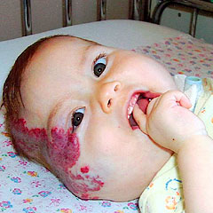
Gemangioma (from Greek. Haima - Blood and Angeon - Vessel) - a benign tumor, developing from the vascular wall, is quite common. Various types of hemangioma make up 25% of all benign tumors and 45% of all soft tumors.
Despite the possibility of self-excretion and stop the growth of hemangioma, followed by disappearance, its further current remains unpredictable.
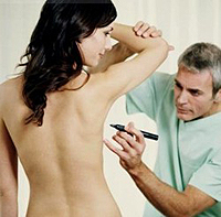
 The diagnosis of hemangioma is carried out by a surgeon doctor using the following techniques:
- inspection;
- laboratory diagnostics;
- Ultrasound examination (determination of the depth of the proliferation of the tumor and the calculation of the volume of education; the features of the location, the structure of the tumor, the measurement of the blood flow rate in the vessels is determined);
- Angiography (obligatory when examining patients with extensive and deep hemangiomas).
Doctor hemangioma
Surgical method is the leading. It is especially used in the localization of the tumor on the skin and in the subcutaneous tissue, as well as in the muscles and internal organs (liver, intestines). The following operations are used: the excision of the tumor, partially removing it and swinging with the firmware of the vessels.
The easiest and most radical way - the excision of the tumor. In order to avoid recurrence, it should be carried out within healthy tissues. In addition, excision with the leaving of a part of the vascular tumor may entail strong bleeding. The latter even in a small amount is most dangerous in childhood. Partial excision of the tumor applies in cases where it is impossible to single-stage radical removal. The remaining parts of the tumor can be removed during repeated interventions, as well as with the use of cryotherapy. The skin defects formed after the excision of the hemangioma are closed with a change of free skin flap or apply other types of skin plastics.
Slugging with the firmware of vascular tissue through the skin is a very old method used to reduce blood flow to tumor and obliteration of vascular cavities. This technique is applied with large cavernous hemangiomas, it gives an infidental medical and cosmetic effect, and now it has almost completely refused. Properly performed tumor removal leads to full recovery. The emergence of recurrences indicates incomplete removal of tumor elements, and not on the malignant.
Cryotherapy - Treatment with snow of carbon dioxide - is used with a small size of the skin hemangiome, to remove the tumor sections remaining after operation and during its relapses. This technique is based on the development of aseptic inflammation, followed by vessel tissue tissue. Cryotherapy holds a doctor.
Technique. From the cylinder with carbon dioxide, a snow mass temperature is gained into a leather bag - 78-80°WITH. A piece of snow wrapped in gauze tightly pressed to the surface of the tumor at 15-30. Repeated applications can be made in 12-4 days after the sacrifice of inflammatory phenomena, just up to 5 times.
70 are used as a sclerotic substance° ethyl alcohol, which is injected under the tumor and in the circumference of it. Repeated injections of 0.5-5 ml after 7 -10 days can cause vessel obliteration. Sclerosing solution cannot be administered intraderially to avoid skin necrosis. Injections are pretty painful, treatment takes a long time, he has a surgeon.
In the hemangiome of small sizes, the doctor applies electrocoagulation. The method is based on tissue destruction when passing the current of high frequency, it is quite simple, but painful and therefore requires local anesthesia, especially in children. The procedure can be repeated after 7 - 10 days.
Radiation therapy as an independent method is shown at a cavernous, cavernous capillary and capillary hemangiome, when it is localized mainly on the face, where all other methods lead to poor cosmetic results. X-rays and radioactive radiation of cobalt and cesium preparations are used. For the treatment of capillary hemangioma use appliques with radioactive phosphorus and strontium. In children, radiation therapy is rarely used.
The technique is to apply external irradiation by appliqués of radioactive preparations or a near-focus X-ray. The exposure field is strictly limited to the sizes of the tumor. Treatment is carried out by a fractionally one course. The total radiation dose in adults 3500-4000 is happy, in children 2000-2500 Rad. Duration of irradiation 5-6 hours per day (in young children 3-4 h). Daily dose 300-400 Rad. When applying x-ray therapy, one-time dose 250-500 is glad. The intervals between X-ray therapy sessions are 1-2 months. Total dose in adults 2500-3000 Rad, children are 1500-2000.
The best results of the treatment of hemangioma are achieved by excision of a tumor in early childhood with small sizes.