None of us is insured against injuries and especially children who are spent most of the time in active movement. For what signs you can assume the presence of a fracture at the kid? What first help you need to render? How will the treatment take place and how soon the baby will be able to fully play?
Content
Features of bone fractures in children
The bones of the child contains a greater number of organic substances (protein of Ossein) than in adults. Sheath covering bone outside (periosteum) fat, good bloods1. Also, children have a bone tank growth zones. All these factors determine the specifics of children's fractures.
 Often fractures of bones in children occur by type «Green branch». Externally, it looks as if the bone was pulled and bent. At the same time, the displacement of bone fragments is insignificant, the bone breaks only on one side, and on the other side the thick odds holds bone fragments.
 - The fracture line often passes through the growth zone of bone tissue, which is located near the joints. Damage to the growth zone can lead to its premature closure and subsequently to the formation of curvature, shortening, or a combination of these defects in the process of growth of the child. Than at an earlier age, the growth zone occurs, the more difficult consequences it leads.
- In children more often than adults, bone growing fractures arise to which muscles are attached. Essentially, these fractures are torn ligaments and muscles with bone fragments from the bone.
- Bone fabrics in children are faster than adults, which is due to good blood supply to enemotes and accelerated bone corn formation processes.
- Children of the youngest and middle age groups are possible self-correction of residual displacements of bone fragments after a fracture, which is associated with the increasing bone and muscle functioning. At the same time, alone offsets are subject to self-correction, and others. Knowledge of these patterns is important for solving the issue of surgical treatment of fractures.
Types of fractures
Depending on the state of bone tissue, traumatic and pathological fractures distinguish. Traumatic fractures arise from exposure to unchanged bone of short-term, significant mechanical power. Pathological fractures arise as a result of certain painful processes in the bones that violate its structure, strength, integrity and continuity. For the occurrence of pathological fractures of a rather minor mechanical impact. Often, pathological fractures are called spontaneous.
Depending on the state of the skin, fractures are divided into closed and open. With closed fractures, the integrity of the skin is not broken, bone fragments and the whole fracture area remains isolated from the external environment. All closed fractures are considered to be aseptic, uninfective (unimpressed). With open fractures, there is a violation of the integrity of the skin. The dimensions and nature of damage to the skin differ from point wound to a huge defect of soft tissues with their destruction, disintegration and contamination. Special view of open fractures are firearms. All open fractures are primary infected, t.E. having microbial pollution!
Depending on the degree of disagreement of bone fragments, fractures are distinguished without offset and displaced. Fractures with displacement can be complete when the bond between bone frails is broken and there is a complete disagreement. Incomplete fractures when the relationship between fragments is broken not all over, the kost's object is more preserved or bone fragments are held by periosteum.
Depending on the direction of the fracture line, the longitudinal, transverse, oblique, screw-shaped, star, T-shaped, V-shaped fractures with cracking bone.
Depending on the type of bones, fractures of flat, spongy and tubular bones are distinguished. Flat bones include skull bones, blade, iliac bones (form pelvis). Most often, with fractures of flat bones of a significant displacement of bone fragments, it is not. Sponge bones include vertebrae, heel, tranny and other bones. The fractures of spongy bones are characterized by compression (compression) of bone tissue and lead to bone squeezing (decrease in its height). Tubular include bones forming the basis of the limbs. Fractures of tubular bones are characterized by a pronounced displacement. Depending on the location of fractures of tubular bones, they are diaphysar (fracture of the middle part of the bone - diaphysis), epiphyseal (fracture of one of the ends - epiphyse, as a rule, coated with articular cartilage), metaphizar (fracture of the part of the bone - methyphys located between the diaphysia and epiphysis).
Depending on the number of damaged areas (segments) of the limbs or other organism systems, there are isolated (fractures of bones of one segment), multiple (bone fractures of two or more segments), combined (bone fractures in combination with cranial trauma, injury of abdominal organs or chest).
Suspect the presence of a fracture in a child is easy. Most often the child is excited, crying. The main symptoms of bone fractures in children are pronounced pain, swelling, swelling, deformation of the damaged limb segment, the impossibility of the function (for example, the impossibility of moving the hand, step on the leg). Blooding (hematoma) can develop in the zone of projection of the fracture.
A special group of fractures in children constitute compression fractures of the spine, which occur in atypical injury, as a rule, when falling on the back with a small height. The cunning of these fractures is that their diagnosis in children is difficult even with hospitalization in traumatological departments of children's hospitals. Painbacks in the back are insignificant and completely disappear in the first 5 - 7 days. X-ray study does not always allow to put the correct diagnosis. The difficulties of the diagnosis of this group of fractures are associated with the fact that the main radiological sign of the vertebral damage as a result of injury is its wedge-shaped form that children are a normal feature of the growing vertebra. Currently, modern methods of radiation diagnostics are becoming increasingly important in the diagnosis of compression fractures of vertebrals - computer and magnetic resonance tomography.
Bone fractures The pelvis belongs to severe damage and manifest themselves pronounced pain, the inability to get on their feet, swelling and deformation in the pelvic area, sometimes creaping (crunch, creak) of bone fragments when driving with legs.
First aid
First aid for fractures of the limbs is to immobilize, damaged segment with the help of submitted means (plate, stick and other similar objects), which are fixed by a bandage, handkerchief, scarf, piece of fabric and t.NS. In this case, it is necessary to immobilize not only the damaged area, but also two adjacent joints.. For example: when fractures of the bones of the forearm, it is necessary to fix the damaged segment of the limb and the rayscass and elbow joints, during the fractures of the leg bones - the damaged segment of the limb along with the knee and the ankle joints.
To remove the painful syndrome, the affected can be given an anesthetic based on paracetamol or ibuprofen. It should be tried to calm the child, above all, with their calm behavior. Then call
The damaged section of the skin is freed from the clothes (the hands of the one who assisted the help must be washed or treated with alcoholic solution). In arterial bleeding (bright red blood flows over a pulsating jet) It is necessary to press the bleeding vessel above the place of bleeding - where there are no large muscle masses, where the arterie is not very deep and can be attached to the bone, for example, for the shoulder artery - in the elbow. In venous bleeding (the blood of the dark color is poured continuously and evenly, does not pulsate) it is necessary to press the bleeding vein below the place of bleeding and fix the damaged limb in the raised position.
If bleeding does not stop, close the wound with a large piece of gauze, clean diaper, a towel, hygienic gasket (clamping the wound should be checked until the doctor arrives).
 If there is no bleeding with a fracture, then the dirt, scraps, land should be removed from the skin surface. Wound can be rinsed under the jet of running water or pouring hydrogen peroxide (the resulting foam must be removed from the edges of the wound with a sterile marlevary napkin). Next on the wound should be imposed sterile dry bandage. Open fracture is an indication for vaccination against tetanus (if it was not previously conducted or expired since the last revaccination), what needs to be done in an injury or hospital.
The first assistance in falling from the height is to immobilize the spine and the pelvis, which are often damaged. The victim must be put on a solid smooth surface - shield, boards, hard stretchers and t.D. In suspected of a fracture of the bones of the pelvis in the populated areas of the legs, roller fit. All this leads to muscle relaxation and prevents the secondary displacement of bone fragments.
If the child has a damaged hand and it can independently move to go to a children's traumatology point, which, as a rule, is with every children's clinic and hospital.
If the child has a feet injured, spine or pelvic bones, then he cannot move independently. In these cases, it is advisable to call an ambulance aid, which will deliver the affected child to the adoptive department of the children's hospital.
Hospitalization in the hospital is carried out in cases of bone fractures with displacement requiring reposctions (fragments of fragments) or operation, as well as with fractures of the spine and pelvis.
Diagnosis of bone fractures in children is carried out in racecakes or receiving departments of children's hospitals by traumatologists or surgeons. Of great importance for the proper formulation of the diagnosis has a medical examination, a poll of parents, witnesses or a child about the circumstances of injury. Definitely conduct x-ray. Also often (especially when suspected of a spinal fracture) produce computer or magnetic resonance tomography. In the case of a combined injury to diagnose the state of the internal organs, ultrasound studies (ultrasound) are carried out, blood tests, urine and t.NS.
Treatment
In connection with a fairly fast battle of bones in children, especially under the age of 7, the leading method of treatment of fractures is conservative. Fractures without bone fragment displacement are treated by imposing gypsum Longets (an option for a gypsum bandage, covering not the entire circumference of the limb, but only part of it). As a rule, the fractures of bones without displacement are treated outpatient and do not require hospitalization. Outpatient treatment is carried out under the control of the traumatologist. The frequency of visiting a doctor under the normal course of the fire of the fracture is 1 time in 5 - 7 days. Criterion for properly superimposed gypsum bandage is an element of pain, no impairment of sensitivity and movements in the fingers of the brush or foot.
When fractures with displacement, with severe convolver, intra-articular fractures, an operation under general anesthesia is carried out - a closed reposition of bone fragments with the subsequent imposition of a gypsum bandage. Duration of surgical manipulation - a few minutes. However, the conduct of anesthesia does not allow to immediately release the child home. The victim should be left in the hospital for several days under the supervision of the doctor.
With unstable fractures for the prevention of the secondary displacement of bone fragments, proboscitimate fixation with metal knitting needles is often used, t.E. Bone fragments fixed with knitting needles and additionally gypsum bandage. As a rule, the method of reposition and fixing the doctor determines before manipulation. When fixing the area of fracture with the knitting needles, leaving and gleaming of the spokes out of the limb are needed, such a method provides reliable fixation of the fracture and after 3 - 5 days a child can be discharged on outpatient treatment.
In child traumatology, the method of constant skeletal stretching is widely used, which is most often used in fractures of the lower extremities and is to carry out the needles through the heel bone or the tibia bone (lower leg bone) and the extraction of the limb by cargo for the battle time of the fracture. This method is simple and efficient, but requires inpatient treatment and permanent observation by a doctor to full fracture.
Recovery period
The deadlines for the battle of fractures in children depend on the age of the patient, the location and nature of the fracture. On average, the fractures of the upper limb will grow on time from 1 to 1.5 months, fractures of the bones of the lower limb - from 1.5 to 2.5 months from the moment of injury, fractures of the bones of the pelvis - from 2 to 3 months. Treatment and rehabilitation of spinal compression fractures depend on the age of the child and can last up to 1 year.
Of particular importance for fracturing in children has rational food. In this regard, it is advisable to include in the treatment regimen of vitamin and mineral complexes containing all groups of vitamins and calcium.
With severe open fractures complicated by circulatory disorder, treatment with oxygen under increased pressure in the barocamera is a hyperbaric oxygenation method (used to prevent infection and contributes to the activation of metabolic processes in the body).
Recovery treatment (rehabilitation) begins in the hospital, and then continues on outpatient conditions. With severe injuries, accompanied by a pronounced violation of the function of the damaged segment, treatment is carried out in rehabilitation centers, as well as sanatorium-resort treatment.
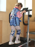
 The active recovery period begins after removing gypsum immobilization or other types of fixation. Its purpose is to develop movements in adjacent joints, strengthening muscles, restoring the supporting ability of the injured limb, etc. Recovery Treatment Treatments include Medical Physical Culture (LFC), Massage, Physiotherapy, Swimming Pool. Physiotherapy and massage are carried out by courses 10 - 12 sessions and contributes to improved blood microcirculation and lymph in a damaged area, restoration of muscle function and joints.
Complications of fractures
In case of complex fractures, a pronounced violation of the function of the damaged limb, pain syndrome. Open fractures are often accompanied by circulatory disorders. The consequences of undiagnosed compression spinal fractures in children lead to the development of youth osteochondrosis - dystrophic (associated tissue disorder) of the spine, in which the intervertebral discs are affected, which is accompanied by their deformation, a change in height, stratification. Also such fractures can lead to spinal deformations, posture disorders and resistant pain syndrome. Bone fractures The pelvis may be accompanied by damage to hollow organs, for example, bladder.

