After examining the inner surface of the intestinal tube with the help of a special unique tower machine - probe, a doctor can identify serious diseases, among which cancer tumors, nonspecific ulcerative colitis, PC, Diverticula, Crohn's disease. Sometimes colonoscopy is actively used to carry out operations on the colon (its length is about one and a half meters). For example, if a coloncoatologist, during this endoscopic study, notes a polyp, then it can immediately remove it, and then send it to the laboratory for further inspection under the microscope.
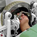
Survey general information

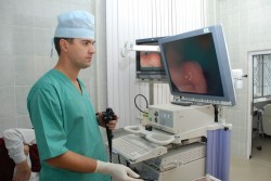
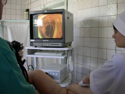
In the course of colonoscopy, as already mentioned, a special probe is involved, called a colonoscope. This is a long flexible tube, equipped with an eyepiece, light source, a device for feeding the air intestine and miniature tongs, allowing to carry out a fence of histological material at the time of the examination. There are currently models of colonoscopes and a built-in camera, it allows you to display an image on a computer monitor (an increased scale allows you to closely consider individual areas and not miss pathological changes), to take photos of the sequined intestinal sites. A colonoscope having several special additional channels is entered directly into the anal hole, and then slowly moving around the entire thick intestine to the place of its transition to the small intestine. On average, the procedure lasts 20-40 minutes. The survey is carried out under local anesthesia or total short-term anesthesia. The doctor has many opportunities during the procedure:
- It estimates the color of the mucous membrane, its glitter, the vessels in the sublifted layer fixes the diameter of the lumen, motor activity, the presence in the intestinal walls of inflammatory processes.
- It can notice different formations on the mucous membrane, for example, polyps, cracks, ulcers, scars, erosion, diverticulus, hemorrhoids, tumors, foreign bodies.
- There is an opportunity with the help of tweezers «bite off» Polyp or a piece of a plot that causes suspicion of pathology. Then all this is passed to the laboratory, where histological studies will be conducted, clarified, is a benign whether the process occurs or a malignant.
- The patient will be delivered from surgical intervention, because the coloncoatologist during the study removes small benign education, polyps.
- When a foreign body is detected, it can be removed.
- If the sector of the intestine is narrowed from abroad, then renovalization is made.
- Manage or drugs eliminate the source of intestinal bleeding.
- You can take a picture of the inner surface of the intestinal tube.
Now in a number of clinics is introduced another new and promising method - virtual colonoscopy. It allows you to get a method of computed tomography image of the walls of the colon. Doctors note the advantages of this method: such a study is less traumatic, it gives more accurate dimensions and localization of areas with pathological changes. But there are also cons: the procedure is expensive, during the survey it is impossible to get tissue samples for histology. In this case, the intestines (to stretch it), air starts, so patients often complain about discomfort in the abdomen and related pain. In order for the colonoscopy to give a doctor as much information as possible, it is necessary to prepare correctly. The MirSovet was already told about this to his readers in the article «How to prepare for colonoscopy».
When colonoscopy is assigned?
There are in medical practice and such cases when it is impossible to conduct this examination:
- With obvious signs Peritonitis;
- during the outbreak of acute infectious processes (regardless of their localization);
- with cardiac and light insufficiency (in late stages);
- in the period when there are signs indicating the aggravation of ulcerative or ischemic colitis;
- If there are problems in the mechanisms of the resulting blood system.
The colonoscopy is not carried out in pregnant women, it is carried out if the patient suddenly begins the attack of the diverticulitis or the Crohn disease is exacerbated.
Whether complications are possible?
Examination with a colonoscope rarely leads to complications. But still we will call them:
- In 1% of patients, the intestines occur - then to restore the damaged zone requires an urgent operation;
- If the patient is under anesthesia, then may (in 05% of patients) there is a drop in pressure, or stop breathing, then resuscitation measures are held urgently;
- It is even less likely to bleeding (both at the time of the procedure and a few hours later and even days);
- If the proctologist removed the polyp, then in the patient in the first few days there may be an increase in body temperature and pain in the abdomen;
- In extremely rare cases after the colonoscopy, it turns out that the patient's infection occurred Hepatitis S or salmonellosis.
If you hit the hands of a qualified, experienced coloncronstologist, then fulfill all its recommendations, be calm during the examination, then it will be successful and will help the doctor to put the correct diagnosis and begin effective therapy.