Arthrosis of the temporomandibular joint - a fairly common disease. The main causes of the disease lies in disadvantages of the dental system. Therefore, precisely the dentists are most often the first to be detected by osteoarthritis.
Content
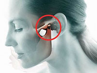 Dull pain, crunch in the area of the lower jaw, limited opening of the mouth — These are typical symptoms of arthrosis of the temporomandulastic joint. Like osteoarthritis of other localization, arthrosis of the mandibular joint is characterized by chronic flow and gradual development of degenerative distofic processes in cartilage and bone tissue. What are the causes of the disease, and what signs indicate the dysfunction of the bone articulation of temporal bone and the lower jaw?
Causes of arthrosis of the temple joint
Osteoarthritis of the temporomandibular joint (ENCH) may be caused by various factors of general and local nature. The total reasons for the disease include metabolic disorders, circulatory innervation in the joint area, hormonal restructuring, affecting metabolic processes, general infectious diseases complicated by arthritis.
The influence of local factors is expressed in an increase in the load on the joint leading to the microtrams of the articular cartilage and their subsequent destruction.
- Bruxism manifested by crossed teeth.
- Lack of indigenous teeth.
- Pathological erasability of teeth.
- Deformed occlusal surfaces of the teeth.
- Non-quality dentures.
- Bribe.
General and local factors are closely related to each other, and complement each other at a certain stage of formation of arthrosis and dysfunction of the mandibular joint. The impetus for the development of the disease can be a violation of exchange in cartilage and bone tissue, leading to a decrease in the adaptive capacity of the joint and damage even under normal load. Bruxism is often combined with a pathological spray of a tooth surface, a decrease in height and deformation of the dentition, which creates unfavorable conditions for the joint.
Pathogenesis of arthrosis of the mandibula
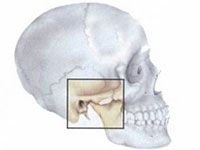
 The development of osteoarthritis ENCH is under the same principles as the arthrosis of another localization. Dystrophic changes are accompanied by a decrease in the ability of the articular cartilage to resist the loads and this leads to its microtrams and destruction. The perforation of the articular disk affects the dumping tissue, causing its restructuring and excessive growth to the formation of osteophytes. The head of the lower jaw sash is deformed, acquires a hooked or pin-shaped, the articular slot is narrowed, the dystrophy of the ligament apparatus and chewing muscles on the side of the defeat. In the presence of local factors creating an excessive burden on the joint, arthrosis of the temple joint develops even faster, bypassing 1 stage.
Arthrosis: Signs of the dysfoliage of the temple joint
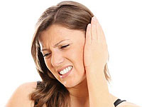
Arthrosis in the field of temporomandibular joints is primarily a new, stupid pain that occurs when the load on this bone articulation. The pain is intensified with chewing, conversation, accompanied by a crunch of various intensity, sometimes click. There is a morning stiffness of the lower jaw, the restoration of the opening of the mouth. Patients often chew food on one side of the tooth row, because the chewing on the other side causes inconvenience and causes pain.
 With an external inspection of the face, the asymmetry of the lower jaw, its fermentation and offset to the side are revealed, nasolabial folds become deep, wrinkles appear in the corners of the mouth. When opening the mouth, it is noticeable as the lower jaw deflects in the sore side or at the very beginning of the movement, or to its end. Further development of Osteoarthrosis of the ENCH leads to a sharp limitation of the opening of the PTA and a violation of normal nutrition.
The diagnosis of the arthrosis of the temporomandibular joint is based on the complaints of the patient, the data of the external inspection, which detects the defects of the dentition and bite, deformation of the face and the joint dysfunction. Detect crunch when moving the lower jaw possible when taking and listening to the joint. With radiography and computed tomography of the joint, the narrowing of the articular gap, degeneration and deformation of the head of the lower jaw, the presence of osteophytes. To diagnose the dysfunction of the joint used registration of movements of the lower jaw, electromyography.

