Dermatomiositis - rare and formidable disease and especially tragic if it develops in children. What are the first symptoms of dermatomyositis, the differential diagnosis of this disease and the main approaches to therapy?
Content
 Dermatomiosit — A rare disease of the connecting and muscle tissue with a motor disorders, redness of the skin, with edema, complicating calcine of soft tissues and purulent infections. The disease is more often celebrated in women, but sometimes found in children.
The reasons for this illness are not studied until the end. The exacerbation of the dermatomyositis causes a long stay in the sun and the presence of infectious agents. Some vaccines against vapor, rubella, measles and typhoids can also be links for the development of dermatomyositis. In this provocateurs of the disease in adults and children are influenza viruses, paragrippa and viral hepatitis in. In addition, bacterial pathogens can be caused, namely, borreliosis and streptococcus group A.
Dermatomiositis: Classification and pathogenesis
The effects of infectious and toxic-allergic factor, burdened heredity, autoimmune shifts play a leading role in the progression of dermatomyositis. The pathogenesis of the disease is based on the occurrence of immune complexes that affect the walls of small blood vessels.
Clinical manifestations of the disease are diverse, so the generally accepted classification divides dermatomyositis by origin and flow.
By origin:
- idiopathic — primary;
- Paranoplastic — secondary;
- dermatomyomy in children;
- in combination with other diseases of the connective tissue.
By the flow of the disease:
- spicy;
- subacute;
- chronic.
Clinical manifestations
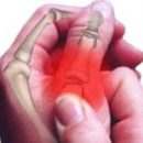
 The main symptoms of the dermatomyositis of adults and children are skin rashes in the form of red and pink spots and peeling plaques in the field of extensive surfaces of the joints. Unlike these skin manifestations, the symptom of the Gottron and the heliotropic rash with juvenile dermatomyositis are sometimes revealed by non-latch blushing nodules.
Leading signs of dermatomyositis, treatment and a differential diagnosis of which is paid to considerable attention, are the defeat of skeletal muscles, lungs, joints, hearts, gastrointestinal tract and endocrine organs.
The lesion of muscles is manifested in the weakness of muscle fibers, which dramatically limits the possibility of performing elementary domestic action. In progression of the disease, not only the pelvic and shoulder muscles can be involved in the process, but the intercostal muscles and muscles of the diaphragm, which in turn can lead to respiratory failure. In addition, inflammatory and degenerative changes in muscle tissue can be the cause of overpressing connective tissue in it, and this contributes to the development of muscle contractures.
Light damage arises due to the weakening of intercostal muscle fibers, which significantly reduces the ventilation function of the lungs, thereby significantly complicating the therapy of dermatomyositis. Interstitial pneumonia and fibrusing alveolitis are leading clinical manifestations of this disease.
Calcine soft tissues seems to be a characteristic feature of dermatomyositis in children. It is based on the deposition of calcium salts in muscles and subcutaneous fatty tissue.
With the defeat of the heart in the pathological process, not only myocardia is involved, but all the shells of the heart are involved, it is manifested by tachycardia and heart rate disorders.
Involvement in the pathological process of the smooth intestinal musculature — This is an active dermatomyosis, the pathogenesis of which consists in violating blood circulation in small vessels of the gastrointestinal mucosa of the gastrointestinal tract and is manifested by gastritis, colitis, ulcerative disease.
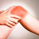
Excessive sprouting of connective tissue in small and large joints is expressed by stiffness, pain and restriction in motion.
Endocrine disorders resulting from the progression of the disease, change the functional activity of the genual glands, adrenal and hypothalamus bark.
Dermatomiositis: Differential Diagnostics
 It is carried out with a red lupus, systemic sclerodermia, mycken, photodermatosis, trichinosis, nodule periatery, exudative erythema, etc.
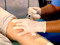
Dermatomiositis, the laboratory differential diagnosis of which is carried out with other skin and systemic diseases based on the conduct of a general, biochemical analysis of blood and histological research.
In general blood test, there is a moderate growth of ESP and a slight leukocytosis in biochemical blood test — Enhance hepatic enzymes. To confirm this diagnosis, a target muscle biopsy is used followed by histological examination.
Dermatomiositis: Treatment
Dermatomiositis involves treatment, which is based on the use of glucocorticoids and cytostatic drugs to maintain the function of the internal organs, as well as drugs that improve blood circulation in the microcirculatory.