What is osteomyelitis? As Osteomyelitis manifests? How to treat osteomyelitis? Answers to these questions you will find in the article.
Content
Osteomyelitis
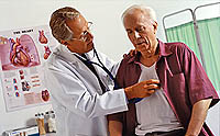
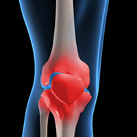
Osteomyelitis - Currently, this title combines the defeat of all parts of the bone: inflammation of the bone (sax), bone marrow (myelitis) and periosteum (periostitis). It arises as a result of endogenous (hematogenic) or exogenous infection in the bone.
The hematogenous osteomyelitis is distinguished, developing as a result of microbes in the bone through blood, and the wound, or traumatic osteomyelitis, which is secondary and develops as a complication of the wound process, operational treatment of closed fractures. In the initial stage, these two types of bone inflammation are completely different both by origin and on manifestations. However, in the later phases, the differences are gradually smoothed, so they wear a common name.
High efficiency of antibiotics significantly improved outcomes in conservative treatment and reduced the need for operations. Treatment of hematogenous osteomyelitis consists of activities of the overall impact on the body of the patient and local - to the focus of infection.
Immobilization conducted from the very first days of the disease contributes to the restriction of the process, reduces pain and improves the overall health of the patient. Operation (it is necessary to resort to it rarely) is shown in running processes with the development of phlegmons in cases where conservative treatment does not succeed, and to remove sequetters.
What is hematogenous osteomyelitis
Hematogenous osteomyelitis - more often occurs in children and teenagers of male. The process is usually localized in femoral and tibial bones and less often in all other. From the primary hearth (furuncula, carbuncula, panaritics, phlegmon, abscesses, face, infected abrasions and raps, carious teeth, tonsillitis, chronic inflammatory processes in the applied cavities of the nose and ear, etc.) Microbes fall into the bone marrow through blood flow and cause inflammation.
The following factors affect the development of osteomyelitis are distinguished:
- anatomy-physiological;
- biological and immunobiological;
- predisposing.
On clinical flow, acute and chronic osteomyelitis are allocated, which in the overwhelming majority of cases is the outcome of the acute, but may occur and as primary chronic.
Symptoms of hematogenic osteomyelitis
In the first 1-2 days of the patient, there is a general ailment, lubrication in limbs, muscle pain, headache. Then an amazing chill appears with a resistant increase in temperature to 39°C, and higher, weakness, breakdown, headache, sometimes vomiting. The general condition becomes heavy, consciousness is darkened, nonsense appears, symptoms of irritation of the brain shells, and sometimes convulsions. Appetite disappears, the language is covered, dry.
The face becomes pale, the eyes are wade, lips and mucous membranes cianotic, dry skin, with a yellow tint, the turgor is reduced. Blood pressure is lowered, the tones of the heart is deaf, the pulse is frequent, weak filling and, as a rule, corresponds to temperature. Student breathing, superficial. Sometimes symptoms of bronchopneumonia are sometimes found in the lungs. Liver and spleen are increased, painful in palpation. Sometimes painful area of kidneys, urine is not enough, in the urine of protein and cylinders.
 With 1-2 days of the disease, there is strictly localized severe pain in the affected limb, bearing the fat, drilling, driving character. Patients, especially children, at the slightest movements of the limb, pushes the bed, shouting often screaming from the amplification of pain. To reduce pain, they lie completely motionless. Due to the deep location of the focus, the methodological palpation is acquired in such cases, which must be carried out carefully. It allows you to designate the area of the greatest pain, the corresponding process center. The method of early diagnostics - the heel harness or on the elbow, which causes severe pain in the place of defeat.
In the next 1-2 days, local phenomena are more distinct. Accordingly, the location of the lesion appears painful swelling of soft tissues, which quickly increases, moderate redness and skin sweering, increase its temperature. Despite the fact that x-ray study during this period does not give any data, the diagnosis becomes quite clear.
Failure of the limbs rapidly increases, begins to shine extended veins, regional lymphatic nodes increase. At the end of 1 weeks in children and weeks in two teenagers in the center of painful and dense swelling, fluctuation begins to determine. With the development of intertensive phlegmon, the overall state of the patient is somewhat improved, if it remains difficult, you need to look for certain complications (transition of the process to the nearby joint, multiple bone damage, the formation of feminic foci, etc.).
Without surgical treatment, the intertensive phlegmon can independently reveal with the subsequent formation of the fistula. In more adverse cases, it progresses and leads to secondary purulent arthritis, paraarticular phlegmon and sepsis.
The course of acute hematogenous osteomyelitis depends on the timeliness of the proceeding, in particular the use of antibiotics. This is indicated by medical practice, noting the increase in recent times «Subacle» forms and significant reduction in the number of acute and septic cases.
What is chronic hematogenous osteomyelitis
The reason for the transition of acute osteomyelitis into chronic is the continuing necrosis of the infected area of the spongy or compact bone layer. The sequester formed is one of the main pathologists of supporting reactive inflammation of the surrounding bone tissue. The weak development of regenerative processes caused by a sharp disruption of the nutrition of the bone and periosteum contributes to chronic flow.
Symptoms occur with the weakening, the body's resistance nesting in the bone infection (injury, cooling, general severe disease, etc.).
X-ray study is valuable to determine the localization and length of the focus of the lesion, helps to establish the nature of the existing pathological changes. The first radiological symptoms begin to be detected from 10-14 days of the disease. In a number of obscure cases, it is advisable to use tomography.
In chronic osteomyelitis flowing to the formation of fistula, the fistulography occupies an important place. It allows you to clarify the localization of the sequestration and reveal when ordinary pictures are not clear enough. In fistulography, contrast agents are used (iodolipol, sergozin, diodon, etc.). The most accurate bone lesion zone can be determined by radioactive scanning using radioactive technetium, which is extremely important for solving the issue of the operation.