The main task of the survey is the identification of current diseases of the body or brain, which could cause epilepsy attacks.
Content
Full medical examination includes a collection
information about the life of the patient, the development of the disease and, most importantly, very
Detailed description Pr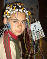 Epilepsy Epilepsy, as well as states, prior,
sick and eyewitnesses of attacks. If the attacks arose in a child,
then the doctor will interest the course of pregnancy and childbirth.
Necessarily a general and neurological examination,
electroencephalography. To special neurological research
Applies nuclear and magnetic and constructive tomography and computer
tomography.
What is electroencephalography (EEG)?
EEG
— This is a completely harmless and painless study. For his
Conduct to the head are applied and fixed on it with
Rubber helmet small electrodes. Electrodes using wires
Connect to the electroencephalograph, which enhances 100 thousand times
The electrical signals of the cerebral cells obtained from them,
writes them on paper or introduces readings into a computer.
Patient V
The study time lies - or sits in a convenient diagnostic chair,
Being relaxed, with closed eyes. Usually when removing the EEG
The so-called functional tests are carried out (photostimulation and
hyperventilation), which are provocative brain loads
through bright light flashes and reinforced respiratory
Activity.
If during the EEG started the attack (which happens very rarely),
then the quality of the survey is much increasing, because in this case
It is possible to more accurately establish an area of disturbed electrical
Brain Activity.
Many changes to EEG
are nonspecific and represent only auxiliary
Information for epileptologist. Only on the basis of the identified changes
the electrical activity of the cells of the brain can not talk about epilepsy, and,
on the contrary, it is impossible to exclude this diagnosis with normal EEG if they have
Place epileptic attacks. Epileptic Activity for EEG
regularly detected only in 20–30% of people with epilepsy.
Necessary
Bill that the interpretation of changes in bioelectric
Brain Activity — it's in some extent art. Changes, related to
on epileptic activity, may be caused by eye movement,
swallowing, pulsation of blood vessels, breathing, movement of the electrode,
Electrostatic discharge and other venerans.
except
that electroencephalography should take into account the age of the patient, since
EEG of children and adolescents is significantly different from the electroencephalogram
Adults.
What is a trial with hyperventilation?
This is
Frequent and deep breathing for 1–3 minutes. Hyperventilation causes
pronounced exchange changes in the substance of the brain due to intense
removal of carbon dioxide (alkalosis), which, in turn, contribute
The emergence of epileptic activity on EEG in people with attacks.
Hyperventilation during EEG recording allows you to identify hidden
epileptic changes and ut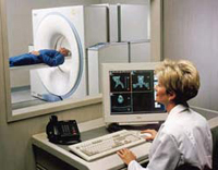 Find the nature of epileptic attacks.
What is EEG with photostimulation?
This
The sample is based on the fact that light flashes in some people with
epilepsy can cause attacks. During EEG recording before the eyes of the patient's examined rhythmic (10–20 times per second) flashes bright
light. Detection of epileptic activity during photostimulation
(photosensitive epileptic activity) allows the doctor to choose
the most correct tactics of treatment.
What is the EEG with sleep deprivation?
Deferre
(deprivation) sleep for 24–48 hours before EEG is carried out to identify
Hidden epileptic activity in cases complex for recognition
Epilepsy. Sleep deprivation is quite strong provoking attacks
factor. It should be used this sample only under the guidance of the experienced
Doctor.
What is EEG in a dream?
How
It is known for certain forms of epilepsy changes to the EEG stronger
expressed, and sometimes only capable of being caught during
Research in SN. EEG recording during sleep allows you to detect
epileptic activity of most of those patients who in
daytime she was not detected even under the influence of ordinary
provocative samples.
But, unfortunately, for a similar study
Special conditions and preparedness of medical
personnel, which limits the wide use of this method. Especially
It is difficult to carry out children.
CT scan
Computer
Tomography (CT) — This is a method of studying the brain using radioactive
(X-ray) radiation. During the study, a series is held
Brain pictures in various planes, which allows, unlike
ordinary radiography, get the image of the brain in three dimensions.
CT allows you to identify structural changes in the brain (tumor,
Calcifications, atrophy, hydrocephalius, cysts, etc.).
However, data CT
may not have informative significance in certain types of attacks,
To which include, in particular: any epileptic attacks in
For a long time, especially in children; generalized
epileptic attacks with the lack of focal changes on EEG and
Instructions for brain damage in neurological examination.
Magnetic resonance imaging
Magnetic resonance
Tomography is one of the most accurate diagnostic methods
Structural changes of the brain. Nuclear magnetic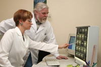 NASS (NMR)
— This is a physical phenomenon based on the properties of some atomic
nuclei when placing them in a strong magnetic field absorb energy in
radiofrequency range and radiate it after termination of exposure
Radio frequency impulse. According to its diagnostic capabilities of NMR
Excellence computed tomography.
To the main disadvantages usually
include: low reliability of identification of calcifications; High
price; the impossibility of examining patients with claustrophobia (fear
closed space), artificial rhythm drivers
(cardiimuels), large metal implants from
non-medical metals.
If a person has
epilepsy attacks stopped, and drugs are not yet canceled, then he
It is recommended to carry out the control total and neurological
Examination at least once every six months. This is especially important
To control the side effects of anti-epileptic drugs.
Usually the condition of the liver, lymphatic nodes, gums, hair,
and laboratory blood tests and liver samples are carried out. except
of that sometimes it is necessary to control the number
Blood-Friends. Neurological examination at
This includes the traditional inspection of the neurologist and the holding of EEG.

