What are benign liver tumors? How to diagnose and treat these tumors? Answers to these questions you will find in the article.
Content
Adenoma liver
The adenoma of the liver is benign tumors arising from hepatic cells or epithelial cells of bile ducts.
Depending on their structure, distinguishes:
 benign hepatoma emanating from hepatic cells;
- a benign cholangioma emanating from the epithelium of bile ducts;
- A benign adenoma mixed structure - hepatocholangioma.
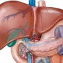
Macroscopically liver adenoma have a kind of round grayish or dark red formations of various sizes located under a capsule or in the thickness of the liver, and may be solid or multiple.
Microscopically, cholangima are divided into a tumor of a solid type, tubular type and cystadema. The adenomes of solid and tubular types usually do not reach large sizes, while cystadenomes can achieve a significant value and are prone to malignant reincarnation.
Hepatoms can be presented in the form of a simple liver dyshambic, encapsulated and clearly rained the structure, or in the form of trabecular adenoma, where the cellular plates are deprived of a lolly location and the tumor does not have its own capsule. The latter are potentially active and prone to malignant reincarnation.
Recently, the adenoma of the liver is increasingly described in women after a long use of oral steroid contraceptives.
Hemangioma liver
Hemangioma liver - the most frequent of all benign tumors of this organ.
Gemangioma refer to vascular tumors. They are benign cavernous blood tumors of the spongy structure (angioma, cavernous hemangiomas and cavities), outgoing from venous liver vessels. Some authors consider hemangiomas not a true tumor, but by defective development, vascular gamartoma.
In the literature, there are instructions that hemangiomas liver are often combined with cysts or cystic lesions of other organs. Hemangiomas liver can be borderless between benign and malignant tumors.
According to the histological structure, there is a cavernous and squirrelic hemangioma. Separately, hemangioendothelima is isolated, in which the signs of non-restrained infiltrative growth characteristic of malignant tumors are detected. By the location of the hemangioma of the liver are superficial, deep and mixed. Hemangiomas liver are found in the form of small multiple formations or single tumors of various sizes.
Nodular hyperplasia
A nodal liver hyperplasia represents a certain oncological interest, since it differs little by its clinical manifestations from the tumors of this organ.
Journal hyperplasia Even during the operation it is difficult to distinguish from the true tumor - cancer or adenoma.
In the occurrence of noded hyperplasia, the local circulatory and biliary disorders in certain sections of the liver.
Macroscopically noded hyperplasias have a kind of dark red, brown or pink formation of various sizes with a smooth or fine-maternal surface. According to consistency, they are denser than intact hepatic cloth, and do not have their own capsule.
Microscopically, with a noded hyperplasia, the picture of the local cirrhosis is found, sometimes very similar to the histological structure hepatom.
It is possible that the local liver hyperplasia is the stage of a single process: local cirrhosis - adenoma - malignant hepatoma.
Non-parasitic cysts
Non-parasitic liver cysts do not represent a big rarity; The reason for their education is different.
Non-parasitic cysts are more often congenital and arise from the adventures of bile ducts and the remainder of the germinal tissue. Traumatic liver cysts are formed from hematoma after leap break.
Cysts of liver ligaments are extremely rare and can be both true, congenital and false - traumatic and inflammatory.
Diagnostics, clinic, treatment of benign liver tumors
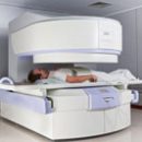
Early diagnosis of adenoma represents a difficult task, since these tumors do not give any pathognomonic symptoms and similar to clinical flow with other benign focal liver diseases. The use of modern research methods when suspected the liver tumor makes it possible to accurately diagnose. The largest diagnostic value is the liver scanning, hepatoogiography and laparoscopy with a sight biopsy.
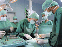
 When clinical signs of tumor and confirmation of the diagnosis of objective research data, the patient must be operated, without waiting for complications. The method of choice in the surgical treatment of the adenoma of the liver is the rescue of the organ within the limits of healthy tissues. A nodal liver hyperplasia for a long time proceeds asymptomatic and manifests itself when large sizes reaches or is detected by chance during other operations.
Features of the clinical course by hemangiom largely depend on their size and distribution.
Asymptomatic liver hemangiomas are clinically nothing manifested, they are found randomly during laparoscopy or on autopsy. As the tumor grows and the increase in its size appears various clinical syptoms - pain and severity in the epigastric region, nausea, air belching.
Among objective symptoms, the presence of a palpable tumor is of paramount importance. Unlike malignant tumors, the hemangioma of the liver is characterized by slow growth, long-term flow and satisfactory condition of the patient.
The clinical course of hemangiom can be complicated by a tumor gap and intra-abdominal bleeding, hemobilia, twisting the leg of the tumor.
Asymptomatic forms of non-parasitic liver cysts are not manifested by anything. As the size of cysts increases, patients make complaints on the feeling of gravity, pressure and pain in the epigastria and the right of hypochondrium.
With an increase in the volume of cyst, the threat of complications is significantly increasing - the breakdown of cysts, suppuration, hemorrhage in the cavity of cysts, mechanical jaundice.
In order to diagnose benign liver tumors: ultrasound and radioisotope examination, computed tomography, angiography, laparoscopy.
Treatment of benign liver tumors surgical. The volume of resection of the liver depends on the size and localization of benign tumors, ranging from segmentectomy and ending with an extended right-sided hemigepatectomy.
With liver cysts, an autopsy and drainage of the cavity of the cyst, the excision of cysts, liver resection, cystegenic anastomoses and monsupialization of cysts.