What is the adenoma liver? What is a nodular hyperplasia? How diagnosed these diseases? Answers to these and other questions you will find in the article.
Content
Adenoma liver
Liver adenoma - rare benign tumor.
- The liver cell adenoma consists of cells resembling liver cells.
- Cystadenoma consists of small proliferating bile ducts lined with an epithelium with an accumulation of mucus and the formation of a cyst.
The first type is more common in women of childbearing age, the second - in men. It is found in the form of one or more nodes drawn from the liver tissue, has a capsule (shell) with a diameter of 1 to 20 cm. When the adenoma is detected in the liver, operational treatment is shown, t.To. With its energetic growth, a tumor break is possible with damage to vessels and bleeding.
Focal formation of the liver. Benign tumors. Hemangioma hemangioma hemangioma liver - a benign tumor, occurs mainly from the venous elements of the liver, usually find accidentally when ultrasound or CT (computed tomography). Possible complications: compression of bile ducts, vessels, gap with abundant bleeding, malignant rebirth. It is necessary to distinguish from metastases, adenoma, lymphangioma, inefficient hyperplasia. Treatment strictly in specialized hospitals.
Nodular hyperplasia
Nodular hyperplasia - rare tumor-like damage to non-citrotic liver; represented by a plurality of nodes with a diameter of 0.1-4.0 cm, liver changes are minimal, sizes are usually within the normal range. It is necessary to distinguish from cirrhosis, liver metastases. For diagnostics use computer tomography (CT) with contrasting amplification or magnetic resonance imaging (MRI).
Considering, the absence of absolutely accurate and unambiguously testify in favor of a benign tumor of signs and laboratory markers, according to most professionals, a consistent, phased diagnostic approach is needed.
Benign formations include postoperative and post-traumatic liver cysts, liver abscesses.
Abscess liver
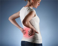
 The liver abscess is a delimited purulent-destructive lesion of the liver, resulting from a drift of infection with hematogenic (with blood current), lymphogenic (with lymph current), cholangogenic (with bile) or contact path. It is more often located in the right lobe of the liver, under the capsule, usually rounded shape and is manifested by discomfort, pain in the right grace and top of the abdomen.
The reason for the occurrence of abscesses is usually intra-abdominal infection.
Liver abscess may also occur after injuries, injuries or operations. Clinical manifestations: fever, pain in the right grace and the right side area, weakness, sweating.
Pretty middle and elderly people are ill. The disease is equally striking both men and women. Clinical manifestations are sufficiently nonspecific and include fever, chills, pain in the right-hand, malaise and weight loss. In 30% of cases, fever may be absent. Almost 45% of patients are presented with complaints about abdominal pain. Many patients prevail the clinical signs of the underlying disease - appendicitis, diverticulitis or biliary lesions.
The most common source of infection (35% of cases) during liver abscesses - biliary tract disease. As a rule, it is cholangitis or acute cholecystitis. In 10-20% of patients with liver abscesses, due to the diseases of the biliary tract, the malignant tumors of the pancreas, the total bile duct and the ampoule of the nipple are detected. Surgical or endoscopic interventions on bile ducts may also lead to the development of liver abscesses. Sometimes liver abscesses are formed due to the parasitic invasion of the biliary tract (round worms or trematodes), which causes the infection of bile. The second frequency source of infection during liver abscesses - intra-abdominal infections, when bacteria fall into the liver on a gate vein. In 30% of cases to the formation of liver abscesses, diverticulitis, krone disease, nonspecific ulcerative colitis and intestinal perforation. Quite rarely the cause of the liver abscess is appendicitis; The exception is the elderly patients and patients with impaired immune status, which are diagnosed with appendicitis late. Approximately 15% of patients with liver abscesses are due to direct penetration of bacteria from a closely located focus of infection, as happens, for example, with subadiaphragmal abscess or with an empiemp of a gallbladder. It is also possible to transfer bacteria into the liver with arterial blood from remote foci of infection (with endocarbage or severe teeth diseases). In 50-70% of cases, pyrogen liver abscesses are caused by gram-negative microorganisms. Most often in such patients found Escherichia Soli, aerobic gram-negative bacterium. Gram-positive aerobes are detected only in 25% of patients; In about 50% of cases, anaerobic microorganisms are causative agents of infection.
Diagnosis of benign tumors
 A ultrasound study (ultrasound) of the liver is always carried out by patients with fever and changed blood test. However, computed tomography (CT) is a more informative method of research to identify destructive changes in the liver.
50-80% of patients with liver abscesses are determined by changes on the overview radiographs of the chest. Symptoms indicating the presence of liver abscesses, serve as atelectasis of the lower share of the right light, pleural effusion on the right and the high standing of the right dome of the diaphragm. The breakthrough of the liver abscess into the pleural cavity can lead to the development of an empieme of pleura. With an overview radiography of the abdominal cavity in 10-20% of cases, air in the abscess cavity is found.
With diagnostic and therapeutic purposes, it is necessary to carry out a puncture fine adult biopsy (PTAB) under the supervision of the ultrasound, which allows you to install a microbial flora, to establish percutaneous clergy drainage in order to conduct therapeutic cavity of the abscess cavity, as well as choose an antibiotic that is the most sensitive to this type of microflora.
Minimally invasive surgical treatment of liver abscesses includes puncture and drainage of its cavity.
Focal formation of the liver. Benign tumors. Minimally invasive surgical treatment of liver abscesses Puncture of abscesses under ultrasound controls with subsequent aspiration of contents, its painting in gram and sowing the nutrient media to determine the sensitivity to antibiotics can help correctly choose antibiotics. Open surgical drainage is performed during the localization of the abscess in the left lobe of the liver and the absence of a noticeable improvement in the state after 24-48 hours after the start of conservative therapy. When localizing an amosh abscess in the left lobe of the liver, the development of complications (tamponade of the heart), accompanied by a high risk of fatal outcome and requiring immediate surgical intervention.
Hematomas and accumulated fluid accumulations - (traumatic, postoperative) accumulation of blood or fluid, which is damaged during damage to vessels in any parenchymal organ or anatomical cavity.
Diagnosed with ultrasound, CT, MRI. The following changes (stages of development) are noted: in the early period, a liquid containing education (clots) is determined; Next, the bunches are converted into the formed masses, partitions of various thicknesses appear, the walls become more dense, thick; With a further increase in fibrous processes and calcination, gradual hematoma resorption occurs; When preserving the liquid component - the formation of pseudokists.