General information about angiography
«Angeion» Translated from Greek means «vessel», but «GRAPHO» - It is literal «Writing, depict». This x-ray study passes with a contrast agent. It allows you to estimate the condition of blood vessels (not only functional), but also to determine the length of pathological changes if they are present. A number of specialists are currently called this study by vasograph. The method is applied not only in radiography, but also in x-ray and computed tomography. And the first time, such a study conducted a scientist in. Forsman in 1929 on their own vessels. Now doctors use two techniques for vessel examination:
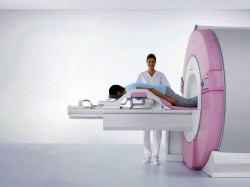
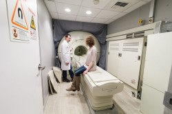
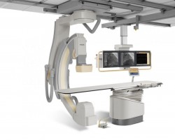
- Invasive - In the patient's artery, a contrast drug is first introduced, which does not harm the body. Then the doctor carefully studies appeared on the monitor «Sleep» internal lumen of the entire branching system of the bloodstream. Thanks to this «Picture» Specialist can judge the bloodstream in a particular organ. The procedure is carried out in specially equipped X-ray operating rooms, there is a high-tech radiological equipment. The devices allow you to make large-format x-rays in a short period of time, in the future they can be subjected to computer processing.
- Non-invasive - designed to study the state of vessels of the lower extremities (or upper), brain, intestines, kidneys, liver and other internal organs to determine the characteristics of the blood flow in them.
Angiography allows you to examine the veins, the arteries of the lower extremities, the light artery, aorta.
When angiography is assigned?
The whole procedure consists of several stages.
First, the patient is introduced the catheter (special tube) into the blood vessel - the radiation artery of the hand (if you need to examine the intestinal vessels, kidneys). If you need to know the state of the heart vessels, the catheter will be introduced directly into the femoral artery. To examine the veins of the lower extremities, the catheter will be introduced into the wreaths in the interfallated intervals. The place of administration is first treated with disinfectants. So that the patient does not have unpleasant sensations, local anesthesia is carried out. Beyond the behavior of the catheter aimed at the vessel, is observed due to x-ray.
Next, the contrast agent is introduced. Then proceed to a quick x-ray shooting. At this time, the patient can experience heat over a few seconds.
Now the catheter is removed, and the prolque place is pressed. This is done in order to stop bleeding. After 20 minutes it will be sterile bandage on the puncture area.
Usually the entire procedure takes no more than 60 minutes. If the doctors after evaluating the state of the vessels and detect problems, they decide to spend immediately balloon angioplasty or other manipulations, the duration of the procedure increases.
Whether complications are possible?
Research is considered safe. And yet, some patients may have:
- Damage to the vessel when puncture and the introduction of the catheter.
- Allergies on a contrast agent (it contains iodine).
After research
Some time (from 6 to 24 hours) After angiography, the patient remains in a medical institution, where medical staff is observing.
We will list a few more important recommendations and prescriptions for past angiography:
- In the first days, drink a lot of fluids so that medicines entered faster from your body will be faster.
- In the first day, relax in bed to bed, so that the body is restarted faster after stress.
- Gradually, you can return to the nutrition recommended by the doctor.
- The imposed bandage should remain in its place for at least 24 hours.
- During the day it is impossible to drive a car and smoke.
- You can take a bath or shower only 12 hours after completion of the procedure.
- You can not lift severity for 48 hours.
Sometimes there feels pain for some time in the puncture of puncture. In the field of the introduction of the catheter may appear. Other complications may occur:
- bleeding;
- hematoma;
- progression of renal failure;
- Cardic impairment, manifestation of heart failure, Myocardial infarction;
- stroke states;
- allergic reactions;
- Vessel injuries.
Carefully watch your condition. Immediately refer to the doctor if:
- Noticed in the field of puncture changes in the color of the skin.
- Hand or leg, where the catheter was injected, hot when touched them.
- The sensitivity of a particular body of the body is lost.
Modern techniques
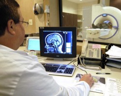
Digital angiography - made of contrasting examination of the vascular system is then exposed to computer processing. As a result, the images of good quality are obtained. And there is an opportunity from the general picture to allocate separate vessels. With this technique, you can not make a puncture to insert the catheter, and the contrast drug to introduce directly into the vein, and the amount of it is significantly less.
Color angiography - the image is made using color coding, helps a specialist to trace the hemodynamics before the start of treatment and after therapy.
3D angiography - images with angiograph, obtained by a digital technique, then exposed to complete the picture of 3D reconstruction.
Angiography allows the doctor to assemble the necessary information about the state of the vessels, to identify their problems (narrowing, malformations, damage, plaques, neoplasms), refine the diagnosis and appoint effective treatment.