The diagnosis of pulmonary artery thromboembolism does not represent difficulties. However, since the manifestations of the disease are not specific, the conduct of primary research is shown to eliminate other pathology.
Content
The presence of risk factors in patient, as well as characteristic features, suggests the presence of an emblem of a light artery. However, manifestations are very nonspecific, therefore, to eliminate other diseases it is necessary to conduct primary research.
Primary survey with pulmonary thromboembolism
 In the overview radiography of the chest organs, with a pulmonal artery thromboembolism, changes are usually not detected. However, this study is absolutely necessary to eliminate other diseases, such as pneumonia, pneumothorax, heart failure, and T.D.
Radiography of the chest organs is necessary to correctly assess the results of scintigraphy of the lungs, carried out by the following stage. Classic wedge-shaped shading detect rarely. More frequent (but non-specific) finds with pulmonary artery thromboembolism: focal infiltration (sealing section), high diaphragm location and pleural effusion.
ECG is often normal or allows you to identify changes related to other diseases. ECG is important to eliminate myocardial infarction. The thromboembolism of the pulmonary artery can determine the appearance of non-specific changes to the ECG. Hypoxia can aggravate myocardine ischemia, in this case the ECG changes can resemble those with ischemic heart disease. With a massive thromboembolism of the pulmonary artery, signs of overloading of the right heart departments are possible.
In arterial blood, the partial oxygen pressure can be reduced during normal or increased partial pressure of carbon dioxide. However, the gas composition of the blood in the embolism of the small branches of the light artery in patients who do not have other diseases may be normal. Thus, normal indicators do not exclude the need for further survey.
Reliable methods of diagnosing disease
The most informative method of diagnosing the thromboembolism of the pulmonary artery is the perfusion scintigraphy of the lungs, which is performed in the first 24 hours of the disease, since sometimes after this term changes disappear.
Angiography of the Liggy Artery - Invasive and not always available diagnostic method. Although it is considered «Gold standard». Angiography is shown in cases where it is necessary to urgently determine the diagnosis, and other research methods turned out to be untenable.
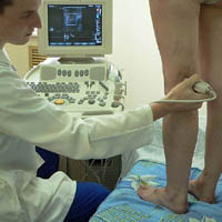
Visualization of the veins of the lower extremities - the study of the first order, regardless of whether the patient has a patient signs of deep vein thrombosis. Detection of deep veins trombosis confirms pulmonary artery thromboembolism.
Compression ultrasound sufficiently makes it possible to estimate the bloodstream in the femoral-pneatheled segment of the lower limb, but often does not reveal asymptomatic clomes in the legs of the lower leg. Since when performing this method, the study does not perform punctures, it can be re.
Echocardiography can confirm the diagnosis of massive thromboembolism of the pulmonary artery, to identify signs of an increase in pressure in the light artery, as well as eliminate other diseases.
Plasma Diameter - Fibrinolysis Process Indicator (Trome Destruction). Diagnostic value has only a negative result of its blood definition. The false-negative result is possible with thromboembolism in the recent past and thromboembolids of the branches of the light artery. This study is used only for additional exclusion of the diagnosis of pulmonary artery thrombosis or lower limb thrombosis.


