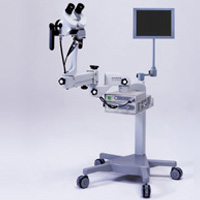
The main methods of diagnosis of leukoplakia are colposcopy and biopsy. Only during biopsy can be reliably diagnosed. The material for biopsy is taken from the focus of the defeat and is sent to further research.
Content
Diagnosis of leukoplakia
The leukoplakia of the cervix - the thickening of the surface layer of the vaginal part of the cervical cervix, is manifested in the form of dry plaques of whitish or yellowish color, generated by an enhanced mucous membrane.
The leukoplak diagnosis does not represent significant difficulties, the foci of the disease is found in the inspection of the genital organs (the area of small sexual lips, the clitoris) and the study with the help of mirrors (cervical area and vagina).
The main purpose of the diagnosis is to determine the nature of the leukoplakia - simple or with manifestations of basal cell hyperactivity and atypics of cells. In case of inspection, there is a particularly alertness against active proliferation and atypics cause leukoplakia with a warthamy surface.
Colposcopy in the diagnosis of leukoplakia
However, the true character of leukoplasts is determined in colposcopy, cytological and histological examination. Colposcopy is mandatory and is reused to exclude or timely recognize attributes atiypics. In this case, the research method is possible to identify other pathological processes that are not observed in case of inspection (inflammatory reaction, flat conclimates, signs of intraepithelial cancer), as well as additional foci of leukoplakia.
Signs of atypics include the following colposcopic paintings leucing. Manifestations of atypics of the transformation zone consider:
- Open withdrawal gradual glands with protruding surfaces by irregular edges
- A large number of vessels, varicose vessels, point vessels around the glands
 In papillary bases, leucing a histological examination reveals a dysplasia or preinvasive cancer.
A comprehensive study required for diagnostics leucing, includes a cytological study. Material for this is obtained by a germ surface of leucoplakia. A cytological study allows you to identify atiypius cells, but atypical cells of the base layer of the epithelium sometimes do not fall into the material, so biopsy is mandatory in all cases of leukoplakia. Biopsy is performed with a sufficiently deep seizure of subepitial tissue. At the same time, they make an excision of leukoplakia foci, if they are small and few.
When leukoplakia is recognized on the cervix, the biopsy is performed with the simultaneous diagnostic scattering of the mucous membrane of the cervical canal.
Timely diagnosis of leukoplakia makes it possible to further avoid such a serious disease as cancer of any localization of female genital organs.


